A significant new brain imaging study is prompting a re-evaluation of one of the most prominent explanations for the persistent and debilitating symptoms of long COVID. Contrary to prevailing theories that suggested widespread inflammation within the brain as a primary driver of these lingering effects, researchers at the University of Turku in Finland have found no substantial evidence of such inflammation in individuals experiencing prolonged post-COVID-19 symptoms. Instead, their findings point towards heightened activity in specific brain regions associated with emotion, stress, and memory as a more likely correlate of symptom severity. This research, published in the Journal of Neurology, offers a crucial shift in understanding a global health challenge that continues to affect millions worldwide.
Unraveling the Mystery of Long COVID’s Neurological Footprint
Long COVID, a complex and often perplexing condition, has emerged as a significant public health concern since the initial waves of the COVID-19 pandemic. While the acute phase of the SARS-CoV-2 infection can be severe, a substantial percentage of individuals report a constellation of symptoms that persist for weeks, months, or even years. These symptoms are diverse and can include profound fatigue, cognitive impairments often referred to as "brain fog," anxiety, depression, sleep disturbances, and a range of other neurological and physical complaints.
The search for the underlying biological mechanisms of long COVID has been a critical focus for the scientific community. Among the leading hypotheses has been the notion that the SARS-CoV-2 virus, or the body’s response to it, triggers ongoing inflammation within the central nervous system. This proposed neuroinflammation was seen as a plausible explanation for the widespread and varied symptoms reported by patients, particularly those affecting cognitive function and mood. However, direct and conclusive evidence to support this widespread inflammation theory has been elusive, leading to ongoing debate and research.
The Finnish Study: A Closer Look at Brain Activity
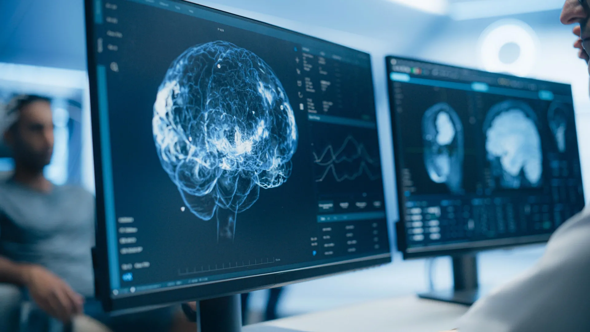
To address this critical question, a team of researchers at the University of Turku, a leading institution in neuroimmunology and part of the InFLAMES Research Flagship initiative, embarked on a comprehensive investigation. Their study utilized cutting-edge brain imaging techniques to meticulously examine the brains of individuals diagnosed with long COVID who continued to experience symptoms well after their initial infection had resolved. The goal was to identify any definitive signs of neuroinflammation and to correlate these findings with the severity and nature of their reported symptoms.
Professor Laura Airas, a leading expert in neuroimmunology and the head of the InFLAMES Research Flagship group, spearheaded the study. Her team’s findings were unequivocal. "We did not observe evidence of widespread brain inflammation in patients with long COVID when compared to healthy controls," Professor Airas stated, directly addressing the core hypothesis under scrutiny. This observation marks a significant departure from the prevailing narrative and suggests that the underlying pathology of long COVID may be more nuanced than previously understood.
Comparative Analysis: Long COVID, Healthy Controls, and Multiple Sclerosis
The study’s robust design involved a comparative approach, assessing not only individuals with long COVID but also two crucial control groups: healthy volunteers and patients diagnosed with multiple sclerosis (MS). The inclusion of MS patients was particularly insightful, as MS is a well-established neurological disease characterized by significant and well-documented inflammation within the brain’s white matter. This allowed researchers to benchmark the inflammatory activity observed in long COVID patients against a known inflammatory neurological condition.
The study cohort comprised 14 individuals diagnosed with long COVID, 11 healthy participants serving as a baseline for normal brain function, and 13 individuals with confirmed multiple sclerosis. All participants underwent a battery of advanced neuroimaging and biological analyses. These included Positron Emission Tomography (PET) scans, specifically designed to detect and quantify neuroinflammation by identifying the presence of activated immune cells in the brain. Magnetic Resonance Imaging (MRI) scans were also employed to assess the overall brain structure, including detailed evaluations of white matter integrity, which is often affected by inflammation. Furthermore, blood samples were collected and analyzed for specific biological markers indicative of neuronal damage and the health of the supporting glial cells within the brain.
The results of this comprehensive comparison painted a clear picture. When juxtaposed with the MS patient group, the long COVID participants exhibited substantially lower levels of inflammatory activity within the brain’s white matter. More strikingly, when comparing the long COVID group with the healthy volunteers, researchers found no meaningful differences in the levels of biological markers associated with either brain inflammation or neurodegeneration. This lack of elevated inflammatory markers in long COVID patients, particularly when contrasted with the known inflammatory profile of MS, strongly suggests that persistent, widespread neuroinflammation is not the primary pathological driver of their symptoms.
The Shifting Sands of Inflammation: Temporal Dynamics
While the study largely debunked the notion of widespread persistent inflammation, it did uncover some intriguing temporal patterns that warrant further investigation. Previous neuropathological studies examining the brains of individuals who succumbed to severe acute COVID-19 had indeed reported clear signs of inflammation. The current study’s findings suggest that this inflammation may be more transient and time-dependent than previously assumed.
Researchers observed that participants scanned within approximately 16 months of their initial SARS-CoV-2 infection displayed higher levels of inflammatory activity in their white matter compared to those who had been infected for a longer duration. Professor Airas proposed a potential explanation for this observation: "This may indicate that inflammation is more noticeable during the earlier stages of the disease before gradually decreasing over time." This temporal aspect suggests that while an inflammatory response might occur during the acute phase of COVID-19, it may largely resolve in the majority of individuals, even those who go on to develop long COVID.
Beyond Inflammation: The Role of Emotion and Stress Pathways
Crucially, the Finnish study identified another significant pattern that appears to be more closely linked to the severity of long COVID symptoms. The researchers discovered a notable correlation between higher levels of anxiety and depression, along with a poorer reported quality of life, and increased cellular activity in two key brain regions: the hippocampus and the amygdala.
The hippocampus is a vital structure within the brain’s limbic system, playing a central role in the formation, consolidation, and retrieval of memories. It is also implicated in spatial navigation and learning. The amygdala, on the other hand, is primarily involved in processing emotions, particularly fear and anxiety, and is crucial for emotional regulation and the initiation of stress responses.
The increased activity observed in these emotion- and memory-related areas among patients with more severe symptoms suggests a potential shift in focus for understanding long COVID. The study’s authors posit that "altered activity in these emotion-related areas of the brain could be connected to the severity of symptoms experienced by some people with long COVID." This finding implies that the subjective experiences of distress, cognitive difficulties, and reduced well-being associated with long COVID may stem from dysregulation within neural circuits governing emotional processing and memory rather than from ongoing widespread tissue damage or inflammation.
Implications for Future Treatment Strategies
The findings from the University of Turku study hold profound implications for the future understanding and treatment of long COVID. By challenging the dominant narrative of persistent brain inflammation, this research compels a re-evaluation of therapeutic approaches.
The researchers emphasize that their results contribute to a more refined scientific understanding of long COVID and question the assumption that persistent brain inflammation is the principal cause of prolonged symptoms in every patient. Instead, the study supports the notion that long COVID is a multifaceted condition where inflammatory changes, if present, are likely most pronounced in the immediate aftermath of infection and subsequently diminish.
Long COVID continues to represent a major global health crisis, impacting the lives of millions with symptoms that can profoundly impair daily functioning and quality of life. The current study’s findings suggest that for a significant subset of these patients, treatments focused on managing stress and improving emotional regulation might be more beneficial than therapies solely aimed at reducing inflammation. This could involve a greater emphasis on psychological support, cognitive behavioral therapy, mindfulness techniques, and other interventions that target the brain’s emotional and stress response systems.
Professor Airas underscored the importance of this shift in perspective: "This study highlights the need to continue investigating the complex biological mechanisms underlying long COVID. Understanding these processes is essential for developing targeted treatments." The research team advocates for a move towards personalized medicine, where treatments are tailored to the specific underlying mechanisms driving an individual’s long COVID symptoms. This might involve identifying distinct patient subgroups based on their neurological profiles, potentially leading to more effective and individualized therapeutic interventions.
The InFLAMES Flagship, a collaborative initiative between the University of Turku and Åbo Akademi University in Finland, exemplifies this forward-thinking approach. This program is dedicated to integrating immunology and related research fields with the express aim of developing novel diagnostic tools and personalized medical treatments. As part of Finland’s Research Council’s Flagship Program, InFLAMES is poised to play a pivotal role in deciphering the complexities of various diseases, including long COVID, and translating these discoveries into tangible patient benefits.
The publication of this study in the Journal of Neurology signifies a critical step forward in the scientific community’s journey to unravel the complexities of long COVID. It provides a robust foundation for future research that will undoubtedly explore the intricate interplay between emotional regulation, stress pathways, and the persistent symptoms experienced by so many individuals, ultimately paving the way for more effective and targeted interventions.
