
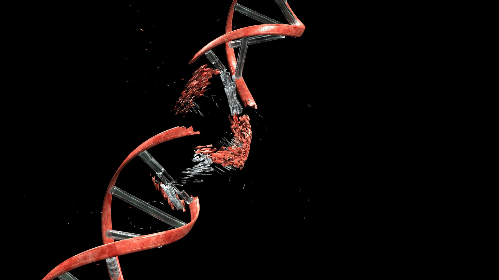
The intricate dance of brain development, a symphony of cellular migration and differentiation, has long been understood as a process fraught with physical challenges for nascent neurons. As these newly formed nerve cells embark on their arduous journeys to their designated locations within the cerebral cortex, they must navigate a dense and complex cellular landscape. This passage, often through tightly packed tissue and narrow interstitial spaces between glial fibers and neighboring cells, was understood to exert considerable physical stress. However, a groundbreaking study published in the prestigious journal Nature has unveiled a profound and previously unacknowledged consequence of this developmental trek: a significant and routine occurrence of DNA double-strand breaks within these migrating neurons.
This revelation, stemming from the meticulous work of researchers at Kyoto University’s Institute for Integrated Cell-Material Sciences (WPI-iCeMS) in collaboration with several other leading academic institutions, challenges conventional understanding of DNA integrity as an absolute prerequisite for healthy cellular function during development. The study posits that these severe forms of DNA damage, where both strands of the DNA double helix are severed, are not necessarily indicators of cellular malfunction or impending doom, but rather an intrinsic, albeit surprising, aspect of normal brain cortex development. Crucially, the research elucidates a sophisticated and highly efficient repair mechanism within these developing neurons that mitigates the potentially catastrophic effects of such damage, allowing the cells to proceed to their final destinations and integrate seamlessly into the brain’s intricate communication network.
The Microscopic Gauntlet: Navigating the Cellular Maze
The journey of a developing neuron is akin to a microscopic odyssey. Born in germinal zones, typically deep within the developing brain, these immature cells must migrate radially or tangentially, often over considerable distances, to populate the layered structure of the cerebral cortex. This migration is a highly orchestrated event, guided by a complex interplay of molecular cues and physical interactions with the surrounding cellular environment. The cerebral cortex, the outermost layer of the brain responsible for higher-level cognitive functions, is characterized by a dense network of neuronal cell bodies, dendrites, axons, and supporting glial cells. As neurons squeeze through this densely packed milieu, they encounter significant physical resistance, forcing them to deform and navigate constrictive pathways.
The Kyoto University-led research team sought to meticulously recreate these physical hurdles in a controlled laboratory setting. By designing and fabricating microfluidic channels, meticulously engineered to mimic the confined spaces and physical pressures experienced by migrating neurons in vivo, the researchers were able to directly observe the cellular response to these developmental challenges. These microchannels, some as narrow as a few micrometers in diameter, served as a sophisticated model system to isolate and study the effects of mechanical stress on neuronal DNA.
The Unveiling of Double-Strand Breaks
Utilizing advanced imaging techniques, specifically the application of fluorescent markers that bind to sites of DNA double-strand breaks, the scientists observed a startling phenomenon. As neurons were guided through these constricted microchannels, the markers illuminated, indicating the formation of double-strand breaks. The intensity and frequency of these fluorescent signals directly correlated with the degree of confinement and the mechanical forces exerted on the cells. This visual evidence provided compelling proof that the physical act of migration through tight spaces was a direct trigger for this severe form of DNA damage.
However, the narrative did not end with the discovery of damage. A critical aspect of the study was the observation of the subsequent cellular response. Once the neurons successfully navigated through the microchannels and emerged into a less restrictive environment, the researchers noted a remarkable phenomenon: the DNA damage began to recede. Within a 24-hour period, the vast majority of these double-strand breaks were efficiently repaired. Crucially, these repaired neurons continued to exhibit normal functionality, integrating into the simulated neural network without apparent impairment. This observation was pivotal, suggesting that the developing brain possesses an inherent resilience to this specific type of DNA damage.
The Culprit: Topoisomerase IIα in Mechanical Stress
The research delved deeper to identify the molecular machinery responsible for this stress-induced DNA damage. The study pinpointed an enzyme known as Topoisomerase IIα (Topo IIα) as the primary instigator. This enzyme plays a crucial role in managing DNA topology, a process essential for numerous cellular activities, including DNA replication, transcription, and recombination. Topo IIα functions by temporarily introducing a double-strand break in the DNA, allowing the DNA to untangle or relieve torsional stress, and then rejoining the broken ends. This mechanism can be conceptually understood as carefully cutting a twisted cable to relieve tension, then splicing it back together.
Under normal cellular conditions, this transient breakage and rejoining is a tightly regulated process. However, the research revealed that when neurons are subjected to significant mechanical stress during migration, the enzyme can become trapped mid-process. The physical forces can impede the rejoining step, leaving the DNA strands broken. In essence, the strenuous passage through constricted spaces can disrupt the enzyme’s ability to complete its task, resulting in persistent double-strand breaks. The cell then relies on a robust DNA repair pathway known as non-homologous end joining (NHEJ) to mend these severed DNA ends, a process critical for restoring genomic integrity.
A Selective Vulnerability: Why Neurons Differ from Cancer Cells
The study further elucidated why this DNA damage, while significant, does not lead to widespread cellular dysfunction or death in developing neurons, a stark contrast to what might be observed in other cell types, such as certain cancer cells. When cancer cells, known for their migratory and invasive properties, were subjected to similar microchannel confinement, the researchers observed a different pattern of DNA damage. In cancer cells, the damage appeared more random and widespread, often affecting critical genomic regions. This indiscriminate damage could disrupt essential gene functions, leading to increased mutations, cellular dysfunction, or even programmed cell death (apoptosis).
In contrast, the DNA breaks observed in developing neurons exhibited a distinct localization. The research indicated that these breaks were predominantly concentrated in genomic regions that are not actively transcribed or involved in the expression of essential genes. This means that the genes critical for neuronal survival and function remained largely intact, even in the presence of transient DNA breaks. This selective vulnerability allowed neurons to tolerate the damage and proceed with their developmental program, a testament to the finely tuned mechanisms that govern brain development.
The Consequences of Incomplete Repair: A Glimpse into Neurological Disorders
While the developing brain demonstrates remarkable resilience, the study also explored the implications of impaired DNA repair mechanisms. To investigate this, the researchers engineered mice whose newly formed cerebellar neurons were genetically modified to lack Ligase 4 (LIG4), a crucial enzyme in the NHEJ pathway responsible for repairing double-strand breaks.
These engineered mice initially appeared to develop normally, showing no overt abnormalities in their early life stages. This observation might suggest that the brain can compensate for some degree of impaired repair during early development. However, as these mice reached adulthood, a subtle yet progressive neurological phenotype began to emerge: mild, worsening balance problems. These motor deficits bear a striking resemblance to symptoms observed in certain human genetic disorders characterized by genome instability, particularly those affecting the cerebellum, a brain region vital for motor control and coordination.
This experimental manipulation provides a crucial link between DNA repair fidelity during neuronal development and long-term neurological health. It suggests that while the developing brain can tolerate a certain level of DNA damage and incomplete repair, chronic or significant failures in these repair pathways can manifest as neurological disorders later in life, particularly impacting motor functions. This finding opens new avenues for understanding the pathogenesis of a range of neurological conditions where genome instability is implicated.
Broader Implications: Brain Diversity and Neurodevelopmental Diseases
The implications of this research extend far beyond the immediate understanding of neuronal migration. The findings suggest that DNA breakage and repair may play a far more significant role in shaping brain biology than previously appreciated. Professor Mineko Kengaku, the lead author of the study and a distinguished professor at WPI-iCeMS, emphasized the paradigm shift this research represents: "It shifts how we think about the neuronal genome. All neurons originate from the same DNA, but DNA damage and repair can introduce small genetic differences between individual neurons through a small mechanical journey. Some of that history may be written into the genome itself."
This suggests that the very journey a neuron undertakes can imbue it with subtle genomic variations. These variations, arising from the dynamic process of DNA breakage and repair, could contribute to the remarkable diversity observed among individual neurons within the brain. While the study did not directly investigate this, it raises the intriguing possibility that these early genomic alterations might influence the unique functional properties and connectivity patterns of individual neurons, ultimately contributing to the complexity and adaptability of the brain.
Furthermore, the link between impaired DNA repair and neurological deficits in the mouse model opens up new avenues for investigating the etiology of neurodevelopmental and neurodegenerative diseases. Conditions such as certain forms of intellectual disability, autism spectrum disorder, and even age-related neurodegenerative diseases like Alzheimer’s and Parkinson’s disease, may have an underlying component related to compromised DNA repair mechanisms during critical developmental windows or throughout life. Future research could explore whether variations in DNA repair efficiency in humans correlate with susceptibility to these conditions or influence their severity.
The collaborative nature of this research, involving esteemed institutions such as the University of Tokyo, the University of Osaka, the National University of Singapore, and the Tokyo Metropolitan Institute of Medical Science, underscores the global effort to unravel the complexities of brain development. This multidisciplinary approach, combining expertise in cell biology, neuroscience, and engineering, has been instrumental in achieving these significant findings.
In conclusion, this seminal study has unveiled a hidden layer of complexity in the intricate process of brain development. The discovery that neuronal migration routinely induces DNA double-strand breaks, and that the brain possesses sophisticated mechanisms to tolerate and repair this damage, fundamentally alters our understanding of neuronal integrity. The research not only sheds light on the remarkable resilience of the developing brain but also provides critical insights into the potential molecular underpinnings of neurological disorders, paving the way for future investigations into brain diversity and the pathogenesis of a wide range of neurological conditions. The very journey of a neuron, it appears, leaves an indelible mark on its genomic landscape, a testament to the dynamic and adaptive nature of the human brain.
