
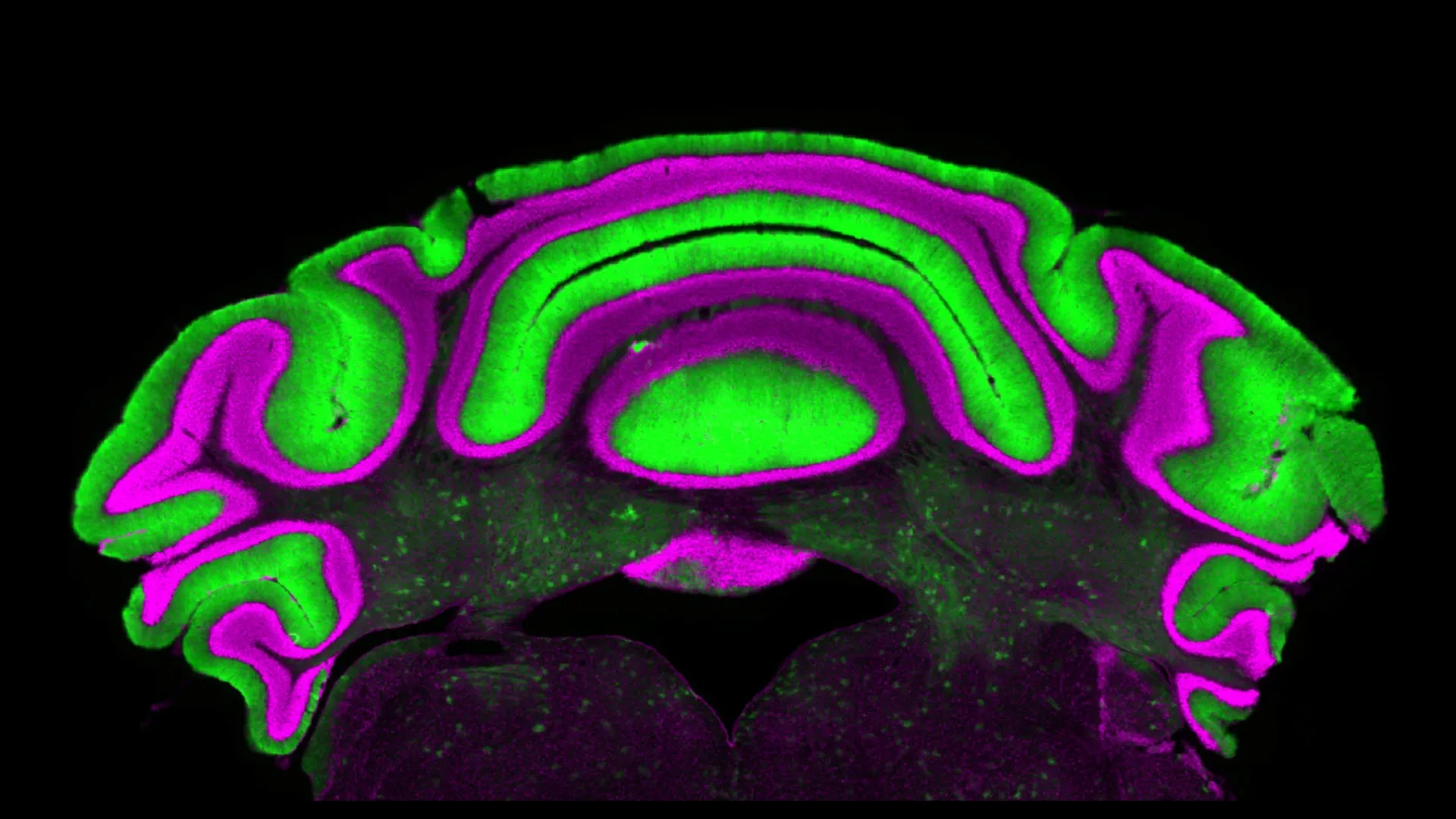
New research from a Virginia Tech neuroscientist at the Fralin Biomedical Research Institute at VTC is raising questions about a long-standing approach to studying chronic neurological conditions such as dystonia, ataxia, and tremor. For decades, the prevailing scientific understanding of how the cerebellum, a crucial brain region for motor control, functions and malfunctions in these debilitating disorders has rested on a foundational assumption: that the activity of one type of neuron, the Purkinje cell, directly and reliably reflects the activity of another, the deep cerebellar nuclei cells. This new study, however, suggests this paradigm may be fundamentally flawed, potentially necessitating a significant shift in how these conditions are investigated and treated.
The Cerebellum’s Central Role in Movement
The cerebellum, a densely folded structure located at the back of the brain, plays an indispensable role in coordinating voluntary movements, maintaining balance, and ensuring smooth, precise motor execution. When the intricate circuitry within the cerebellum is disrupted, individuals can experience a spectrum of movement-related symptoms. Dystonia, characterized by painful and prolonged muscle contractions leading to abnormal postures, can severely impact quality of life. Ataxia manifests as a lack of voluntary coordination of muscle movements, resulting in unsteady gaits and difficulty with fine motor tasks. Tremor, an involuntary and rhythmic shaking, can affect various parts of the body, most commonly the hands. These conditions, often chronic and progressive, represent significant challenges for patients and the medical community alike.
The Purkinje Cell-Deep Cerebellar Nuclei Axis: A Foundational Hypothesis
Historically, neuroscientists have heavily focused on the intricate relationship between two key cell types within the cerebellum: Purkinje cells and deep cerebellar nuclei (DCN) cells. Purkinje cells, known for their elaborate dendritic trees, are the sole output neurons of the cerebellar cortex. Their primary function is to exert an inhibitory influence on the DCN cells, which are the principal output nuclei of the cerebellum, relaying processed motor information to other brain regions. This inhibitory connection has led to a widely accepted assumption: that monitoring the activity of Purkinje cells offers a reliable proxy for understanding the activity of the DCN cells. This assumption has been particularly appealing because Purkinje cells are located in the more superficial layer of the cerebellar cortex, making them relatively more accessible for electrophysiological recordings and other experimental manipulations compared to the deeper-seated DCN cells.
This accessibility has meant that a substantial body of research has centered on analyzing Purkinje cell activity as a primary indicator of cerebellar function and dysfunction. Consequently, many therapeutic strategies and research models have implicitly or explicitly targeted Purkinje cells, with the expectation that changes in their activity would predictably translate to corresponding changes in DCN cell output, and ultimately, motor behavior.
Challenging the Status Quo: The Van der Heijden Study
The new research, spearheaded by Meike van der Heijden, an assistant professor at the Fralin Biomedical Research Institute at VTC, and published in the prestigious Journal of Physiology, presents compelling evidence that this long-standing assumption may not hold true. The study’s findings indicate that the activity of Purkinje cells does not reliably predict the activity of DCN cells, even though a direct anatomical connection exists between them.
"We see that there’s not a clear linear relationship between activity in the Purkinje cells and in the deep nuclei cells," stated Dr. van der Heijden. "So there’s very limited predictive power in monitoring one to understand what’s going on in the other." This observation directly challenges the foundational premise that has guided much of cerebellar research for years.
The research team meticulously analyzed a comprehensive database of electrophysiology recordings. These recordings were collected from pre-clinical models that recapitulated various aspects of cerebellar disease, providing a rich dataset for examining neuronal activity under conditions relevant to human neurological disorders. The analysis focused on simultaneously recorded activity from Purkinje cells and DCN cells.
Unexpected Results from Cerebellar Recordings
Under typical physiological conditions, the inhibitory influence of Purkinje cells on DCN cells dictates a predictable inverse relationship: increased Purkinje cell firing should lead to decreased DCN cell firing, and vice versa. This inhibitory pathway is fundamental to the cerebellum’s ability to fine-tune motor commands. However, when the researchers examined the data from their pre-clinical models of cerebellar disease, they discovered a striking lack of correlation.
"The results revealed no significant correlation between activity in the two cell populations," the study reports. This finding was unexpected and has significant implications. It suggests that the complex network within the cerebellum, particularly in disease states, operates with a level of independence between Purkinje cell output and DCN cell activity that was previously underestimated.
Alyssa Lyon, a doctoral candidate in Virginia Tech’s Translational Biology, Medicine, and Health Graduate Program and the paper’s first author, emphasized the practical consequences of these findings. "Purkinje and cerebellar deep nuclei cell activity is disrupted in a disease state, and a better understanding of the relationship between these neuron types will ultimately help optimize treatments for diseases such as dystonia, ataxia, and tremor," Lyon explained.
Implications for Research and Treatment Strategies
The implications of this study are far-reaching, potentially impacting both the fundamental understanding of cerebellar disorders and the development of future therapeutic interventions.
A Call for Direct Investigation of DCN Cells:
Dr. van der Heijden strongly advocates for a shift in research focus. "If you want to know how the cerebellum is behaving in a disease state, you have to look at the deep nuclei neurons, not just the Purkinje cells," she asserted. This suggests that future research endeavors should prioritize direct measurement and manipulation of DCN cell activity, rather than relying solely on Purkinje cell activity as an indirect indicator. This might involve developing more sophisticated imaging and recording techniques to access these deeper brain structures.
Re-evaluation of Therapeutic Targets:
The findings also serve as a cautionary note for treatment strategies. Many existing and proposed therapies for cerebellar movement disorders aim to modulate Purkinje cell activity. The assumption has been that such modulation would predictably alter DCN output and, consequently, improve motor control. However, if Purkinje cell activity doesn’t reliably predict DCN activity, then targeting Purkinje cells might not yield the desired therapeutic outcomes for the DCN-mediated motor pathways.
"This is a cautionary tale for understanding cerebellar activity in disease, but also for treating these challenging diseases," Dr. van der Heijden commented. "We need to be very careful in making assumptions, and to actually do experiments to test our hypotheses." This underscores the importance of rigorous experimental validation before translating research findings into clinical applications.
Potential for New Therapeutic Avenues:
Conversely, this discovery might also open up new avenues for therapeutic development. If the relationship between Purkinje and DCN cells is more complex than previously understood, it could mean that novel targets within the DCN pathway, or even entirely different cerebellar circuits, could be exploited for therapeutic benefit. Understanding the precise mechanisms by which Purkinje cells do influence DCN cells, and identifying the factors that decouple this relationship in disease states, could lead to more targeted and effective treatments.
Background Context and Chronology
The study’s findings did not emerge in a vacuum. Research into the cerebellum’s role in movement disorders has been ongoing for decades. Early studies, dating back to the mid-20th century, began to elucidate the basic anatomical and functional connections within the cerebellar circuitry. The identification of Purkinje cells as inhibitory interneurons and DCN cells as principal output neurons was a major breakthrough.
The ease of access to Purkinje cells for experimentation, compared to the deeper DCN, naturally led to their prominence in research. For example, optogenetic techniques, which allow for precise control of neuronal activity using light, have been extensively used to manipulate Purkinje cells. This accessibility, coupled with the established inhibitory link, solidified the "Purkinje cell as proxy" hypothesis.
However, as imaging and recording technologies have advanced, particularly in the last two decades, scientists have been able to probe deeper into the brain with greater resolution. This has allowed for more nuanced observations of neuronal activity in complex circuits. The present study, by leveraging advanced analytical techniques on a substantial pre-clinical dataset, represents a critical juncture where these technological advancements are forcing a re-evaluation of long-held assumptions. The timeline of this research suggests a progression from foundational anatomical discoveries to functional studies, and now to a critical re-examination of those functional interpretations in light of new data.
Broader Impact and Implications
The ramifications of this research extend beyond the immediate fields of neuroscience and neurology. It highlights a broader scientific principle: the need for continuous critical evaluation of established paradigms. Scientific progress is often characterized by periods of consensus built upon prevailing theories, followed by moments of disruption when new evidence challenges those theories. This study exemplifies such a disruptive moment for cerebellar research.
For patients suffering from dystonia, ataxia, and tremor, this research offers both a ray of hope and a dose of realism. The hope lies in the potential for more precise and effective treatments that stem from a deeper, more accurate understanding of their conditions. The realism comes from the acknowledgment that current research approaches may need significant recalibration, which could mean a longer road to definitive breakthroughs.
The study’s emphasis on rigorous hypothesis testing and caution against making assumptions is a crucial takeaway for the entire scientific community. It underscores the importance of empirical evidence in driving scientific understanding and therapeutic development. Future research will undoubtedly build upon these findings, aiming to precisely map the complex interplay between Purkinje cells and DCN cells in both health and disease, and to translate this knowledge into tangible benefits for those affected by movement disorders.
The Fralin Biomedical Research Institute at VTC, through this work, positions itself at the forefront of this evolving understanding of cerebellar function. The institute’s commitment to interdisciplinary research and cutting-edge methodologies is likely to be instrumental in navigating these new frontiers in neurological research. As scientists move forward, the focus will increasingly be on directly addressing the activity and dysfunction of the deep cerebellar nuclei, rather than inferring it, potentially unlocking new pathways to alleviating the burden of chronic neurological conditions.
