
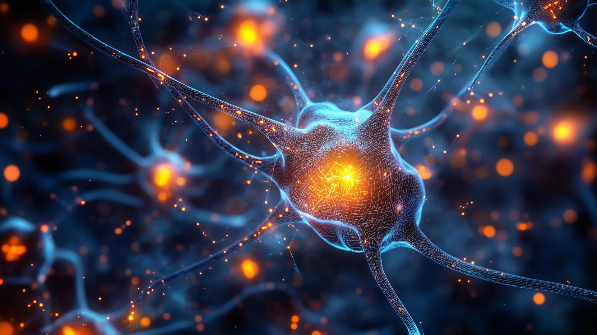
Scientists have identified a group of neurons located in an ancient region of the brain that plays a key role in helping animals focus. These cells appear to improve attention by filtering out distractions and directing the brain toward the most important information. This groundbreaking discovery, made in mice by researchers at Johns Hopkins University, points to a fundamental brain system that is shared across all vertebrates, including humans. The findings, published in the prestigious journal Nature Communications and highlighted as an editorial feature, hold significant promise for developing more precise treatments for attention-related disorders such as Attention-Deficit/Hyperactivity Disorder (ADHD) and autism.
Unraveling the Mystery of Selective Attention
The ability to selectively attend to important information while filtering out distractions is a cornerstone of cognitive function, enabling individuals to navigate complex environments and process vast amounts of sensory input. This skill, known as selective spatial attention, allows humans to engage in conversations amidst the din of a crowded room, locate a familiar face in a bustling throng, or concentrate on a critical task without succumbing to peripheral stimuli. Difficulties in mastering this crucial ability are frequently observed in individuals diagnosed with neurodevelopmental conditions such as autism spectrum disorder and ADHD.
For decades, the prevailing scientific consensus attributed the primary control of attention to the prefrontal cortex, a highly evolved region of the brain that is particularly prominent in humans and other primates. However, this explanation presented a significant evolutionary puzzle. Many animal species, possessing a far less developed prefrontal cortex, exhibit remarkable attentional capabilities. This discrepancy prompted researchers to explore alternative neural mechanisms that might underpin this fundamental cognitive process across the animal kingdom.
"If we really go back in evolution, for hundreds of millions of years, birds have had this ability, fish have had this ability. And they do not typically have a highly developed prefrontal cortex, so how does the brain solve this problem?" explained lead author Ninad Kothari, a postdoctoral fellow in the Department of Psychological and Brain Sciences at Johns Hopkins University. "We were able to identify an evolutionarily old region in the brainstem which affords this ability."
The Brainstem: An Ancient Attentional Engine
The research team at Johns Hopkins University focused their investigation on the brainstem, a primitive part of the brain that is conserved across all vertebrate species. Within this ancient structure, they identified a network of inhibitory neurons that appear to act as a critical filter for sensory information. These neurons, found to be present in species ranging from birds and fish to mammals, were hypothesized to play a vital role in regulating attention.
The impetus for this particular line of inquiry stemmed from prior research conducted by senior author Shreesh Mysore, a neuroscientist specializing in neural circuits tied to behavior, and his colleagues. Their earlier studies on birds, frogs, and turtles had hinted at the involvement of brainstem structures in attentional processes. This foundational work paved the way for the current investigation into the specific function of these neurons in mice.
Experimental Design and Key Findings
To rigorously test their hypothesis, the researchers devised an ingenious experimental paradigm for the mice. This task was designed to mimic the challenges faced by humans when distinguishing between relevant and irrelevant visual stimuli. The mice were presented with visual cues on a screen. Their objective was to correctly respond to information displayed directly in their field of vision while simultaneously ignoring distracting cues that appeared in their peripheral vision. Successful completion of this task was rewarded, reinforcing the desired attentional behavior.
The initial results were striking. The mice performed the attention task with remarkable accuracy, demonstrating their innate ability to focus. However, when the researchers experimentally and temporarily inactivated these specific brainstem neurons, the mice’s performance deteriorated dramatically.
"When we inactivate these neurons, the mice become hyper distractable," Kothari stated, underscoring the immediate and profound impact of disabling this neural circuit. The animals that had previously exhibited focused behavior now struggled to discern between important signals and peripheral noise.
Ruling Out Alternative Explanations
To ensure that the observed deficits were directly attributable to impaired attentional processing, the scientists conducted a series of supplementary experiments. They meticulously ruled out alternative explanations such as vision problems or motor coordination issues. These control experiments confirmed that the mice were not failing the task due to sensory impairment or an inability to physically respond.
Instead, the findings unequivocally pointed to a specific impairment in the animals’ capacity to evaluate competing sources of information and prioritize the most relevant signal. The mice lost the ability to effectively weigh the importance of different visual cues.
"The only thing impaired was their ability to take the competing pieces of information, compare them, and pay attention to the location with the most important information," Professor Mysore elaborated. He further likened this critical brainstem region to an "attentional selection engine," a sophisticated mechanism that continuously answers the fundamental question: "What is the most important information I should pay attention to right now?" This analogy powerfully encapsulates the crucial filtering and prioritization role of these ancient neurons.
Chronology of Discovery and Publication
The journey leading to this significant discovery can be traced back to earlier investigations into avian and amphibian attentional systems. These foundational studies, conducted over several years by Mysore’s lab and collaborators, provided the initial clues that pointed towards the brainstem as a potential key player in attention, irrespective of prefrontal cortex development.
Building upon this groundwork, the Johns Hopkins team embarked on their mouse studies approximately three years ago, focusing on specific neural populations within the brainstem. The experimental design and data collection phase for the core findings of this study spanned roughly two years. Following extensive analysis and validation, the manuscript detailing their discoveries was submitted to Nature Communications. After a rigorous peer-review process, the paper was accepted for publication, a testament to the scientific merit and significance of the research. The publication date of the study was recently confirmed, with its selection as an editorial highlight underscoring its impact within the scientific community. The federal funding for this research played a pivotal role in enabling the comprehensive investigation and advanced experimental methodologies employed.
Broader Implications and Future Directions
The implications of this research extend far beyond understanding basic attentional mechanisms in mice. The discovery of an evolutionarily conserved brainstem circuit responsible for filtering distractions and directing focus opens up exciting avenues for research into human cognition and neurological disorders.
"All the evidence to date suggests that these neurons exist in humans too," stated Professor Mysore, expressing optimism about the translational potential of their findings. "But are they responsible for selective spatial attention in humans? An exciting hypothesis is that they play a crucial role."
The researchers are eager to further explore the precise mechanisms by which these brainstem neurons influence spatial attention across the diverse spectrum of vertebrate species. A critical next step involves investigating whether these neurons perform an analogous function in humans.
The potential impact on the treatment of attention-related disorders is particularly significant. If future studies confirm that these brainstem neurons are indeed crucial for selective attention in humans and that their function is altered in conditions like ADHD and autism, it could revolutionize therapeutic approaches.
"A hallmark of ADHD is that even faint distractors draw attention away — and that’s exactly what we see here when these neurons are silenced," Professor Mysore remarked, drawing a direct parallel between the mouse model and human symptomatology. "But the very next day, when the neurons are turned back on, the same animal can ignore distractors again, even very strong ones." This observation strongly suggests a direct link between the functioning of these neurons and the core difficulties experienced by individuals with ADHD.
Future research will likely involve examining the activity patterns of these brainstem neurons in individuals diagnosed with ADHD and autism. Identifying any functional differences or dysregulations could pave the way for the development of highly targeted medications and therapeutic interventions. Instead of broad-acting treatments, future therapies could aim to precisely modulate the activity of this ancient attentional control system, offering a more nuanced and effective approach to managing these complex conditions.
The collaborative effort behind this research involved a dedicated team of scientists. The authors of the study include Arunima Banerjee, Qingcheng (Jessica) Zhang, and Wen-Kai You, all from Johns Hopkins University, alongside Kothari and Mysore. Their collective expertise and commitment have brought us closer to understanding a fundamental aspect of cognition that impacts both animal behavior and human health. The scientific community eagerly anticipates the follow-up studies that will undoubtedly build upon this remarkable discovery.
