
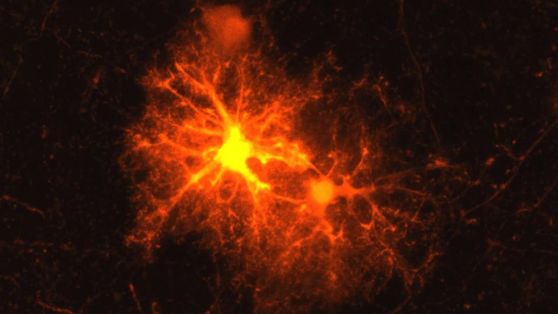
In the complex architecture of the mammalian central nervous system, cellular response to injury has long puzzled neurobiologists. When laboratory mice experience localized brain damage—whether induced experimentally via precise neurotoxin injections or resulting from physical trauma—a predictable cellular cascade unfolds. Experienced neurobiologist Dr. Jan Deussing, a research group leader at the prestigious Max Planck Institute of Psychiatry, had observed this phenomenon countless times over his career: a distinct, highly active population of cells consistently localized to the penumbra of the lesion. Yet, despite repeated observations, the precise identity of these reactive cells remained an elusive biological mystery.
That persistent scientific unknown transformed into an ideal investigative project for Clemens Ries, a master’s student concluding his biology degree, who joined the Max Planck Institute for a targeted research internship. What began as a student thesis evolved into a groundbreaking study that not only identifies a critical player in neural repair but also uncovers an unexpected intersection between stress endocrinology and neurodevelopment. The findings, which redefine our understanding of oligodendrocyte progenitor cells (OPCs) and their newly discovered capacity to synthesize corticotropin-releasing hormone (CRH), were recently published in the esteemed peer-reviewed journal Cell Reports.
Methodical Identification of the Brain’s Versatile Precursors
Tasked with determining the lineage of the mysterious cells clustering around brain wounds, Ries adopted a systematic methodology utilizing advanced murine models. By screening for cell-specific biological markers across the spectrum of known central nervous system cell types, the young researcher narrowed down the possibilities. Only one marker elicited a definitive, robust response: the proteoglycan NG2 and associated markers specific to oligodendrocyte progenitor cells (OPCs).
To fully grasp the significance of this discovery, one must examine the fundamental role of oligodendrocytes within the central nervous system. OPCs are versatile precursor cells whose primary, well-established developmental trajectory involves maturing into oligodendrocytes. These specialized glial cells are responsible for generating the myelin sheath—a rich, lipid-protein multilayered membrane that wraps tightly around neuronal axons. Functionally analogous to the synthetic insulation encasing an electrical copper wire, myelin is indispensable for saltatory conduction, the rapid and energy-efficient transmission of electrical impulses across long neuronal distances. Beyond mere insulation, oligodendrocytes and their progenitor networks provide essential metabolic and trophic support to axons, ensuring overall neuronal survival and homeostasis.
When myelin integrity is compromised, the physiological consequences are profound. In autoimmune pathologies such as multiple sclerosis (MS), the body’s immune system erroneously targets and degrades the myelin sheath, leading to progressive neurological deficits. Similarly, mechanical trauma, ischemic strokes, and neurodegenerative conditions can strip axons of their protective coating, exposing them to degeneration and, ultimately, cell death. Consequently, endogenous remyelination—the restoration of myelin around damaged axons—represents one of the most critical endogenous repair mechanisms available to the mammalian brain following injury.
A Chronological Breakthrough: From Master’s Thesis to Doctoral Discovery
Recognizing the magnitude of initial findings regarding OPC accumulation at injury sites, Ries elected to extend his tenure at the Max Planck Institute of Psychiatry, transitioning the project into a comprehensive doctoral dissertation under Deussing’s mentorship.
Utilizing lineage-tracing techniques and high-resolution confocal microscopy, Ries and his colleagues established a precise chronological timeline of the cellular events following acute brain injury. Within hours of the initial lesion, OPCs residing in the surrounding healthy tissue are activated. These precursor cells undergo a dramatic burst of proliferation, accumulating densely at the margins of the wound. The vast majority of these newly minted OPCs subsequently embark on a differentiation program, maturing into fully functional myelin-producing oligodendrocytes tasked with repairing the structural breach.
However, the research team stumbled upon an entirely unanticipated biochemical feature during this regenerative window. Approximately one-third of the reactive OPCs located immediately adjacent to the damaged tissue began expressing corticotropin-releasing hormone (CRH). Historically, CRH has been understood primarily as a master hypothalamic neuropeptide orchestrating the body’s systemic response to environmental stress through the hypothalamic-pituitary-adrenal (HPA) axis. The revelation that glial progenitor cells—traditionally viewed as passive structural and nutritional support units—could independently synthesize and secrete a primary stress neuropeptide represented a paradigm shift in neuroendocrinology.
The temporal dynamics of this CRH production further underscored its functional importance. The hormonal surge is remarkably acute: CRH expression is detectable within hours of the traumatic event, peaks rapidly, and subsides entirely within approximately three days. This tightly regulated, transient burst strongly implied that glial-derived CRH serves as an immediate early-response signal governing the initial phases of the neural healing cascade.
CRH Receptor 1 and the Fine-Tuning of Remyelination
To decipher the functional role of this injury-induced CRH release, the researchers investigated its downstream molecular targets. The brain utilizes specific receptors to transduce CRH signaling, predominantly CRH receptor 1 (CRHR1) and CRH receptor 2. Through receptor-mapping assays, the team discovered that CRHR1 is constitutively expressed on a distinct subpopulation of OPCs, positioning these cells to directly detect and respond to the extracellular CRH released by their neighboring counterparts.
To test the physiological necessity of this signaling axis, the researchers analyzed genetically modified mouse models lacking the gene encoding CRHR1. The results revealed a complex regulatory mechanism. In the absence of CRHR1, injured OPCs exhibited an exaggerated proliferative response, multiplying at significantly higher rates compared to wild-type controls.
Counterintuitively, however, this hyper-proliferation did not translate into enhanced tissue repair. Longitudinal analysis demonstrated that despite the initial surge in cell numbers, fewer OPCs successfully completed the maturation process into myelin-producing oligodendrocytes. Consequently, the ultimate structural recovery—measured by the density and integrity of the newly formed myelin sheaths—was significantly impaired in mutant mice lacking functional CRHR1 signaling.
This paradox led Deussing and Ries to conclude that CRH does not merely stimulate repair; rather, it acts as a precise temporal and developmental brake. By modulating the pace of OPC proliferation and dictating the precise timing of their differentiation, CRH ensures that progenitor cells do not multiply prematurely at the expense of terminal maturation. Proper timing is absolute; without it, the brain fails to marshal the correct ratio of mature oligodendrocytes required to effectively reconstitute damaged axonal insulation.
Implications for Normal Brain Development and Structural Maturation
The discovery that CRHR1 governs OPC dynamics during injury prompted the research team to investigate whether this same receptor system plays an analogous role during typical, non-pathological neurodevelopment. In humans and rodents alike, myelination is not complete at birth; rather, it is a protracted developmental process that initiates prenatally, accelerates postnatally, and continues progressively throughout adolescence and early adulthood.
Given that uninjured OPCs constitutively express CRHR1 during these developmental windows, the scientists hypothesized that endogenous CRH signaling might shape the baseline architecture of the brain. By examining the brains of mice lacking CRHR1 across various developmental stages using advanced electron microscopy and immunohistochemistry, the researchers mapped structural alterations that persisted well into adulthood.
Mice deficient in CRHR1 displayed heightened OPC proliferation during early postnatal development. Unlike transient injury responses, these developmental alterations became hardwired into the neural architecture. Adult knockout mice exhibited distinct structural anomalies in white matter tracts, notably characterized by aberrantly thick myelin sheaths, particularly surrounding small-caliber axons. These findings confirm that the CRH-CRHR1 axis is a fundamental developmental regulator, dictating baseline myelination patterns and ensuring that axonal insulation scales appropriately with neuronal growth.
Unresolved Questions: The Source of Developmental CRH
While the source of CRH following acute brain trauma is definitively established as the reactive OPCs themselves, normal brain development presents an unresolved biological question. If OPCs synthesize CRH in response to physical injury, what drives CRH signaling during the healthy, uninjured maturation of the neonatal brain?
Based on concurrent neurobiological literature and preliminary mapping data, Deussing and Ries hypothesize that developing neurons fulfill this role. Neurons are well-documented sources of physiological CRH release, particularly under conditions of heightened neuronal activity or environmental stimulation. The working hypothesis posits that as neuronal circuits form and mature, they secrete baseline levels of CRH into the local microenvironment. This neuronal CRH then diffuses to adjacent OPCs, binding to CRHR1 to orchestrate the proliferation-differentiation balance necessary for harmonious myelination.
Broader Clinical Implications: Bridging Neurobiology and Mental Health
The intersection of glial biology, stress endocrinology, and neurodevelopment opens compelling avenues for psychiatric and neurological research. CRH is historically recognized as a central mediator of the neuroendocrine stress response. Furthermore, clinical data consistently demonstrates that early-life adversity and chronic psychological stress are significant environmental risk factors for the development of major depressive disorder, anxiety, and other stress-associated psychiatric conditions.
Pathological analyses of post-mortem psychiatric patient brains have frequently revealed subtle yet consistent alterations in white matter integrity and oligodendrocyte density, suggesting that glial dysfunction may contribute to the pathophysiology of mood disorders. The discovery that the canonical stress hormone CRH directly regulates OPC proliferation and myelination provides a mechanistic bridge linking psychological stress to structural brain remodeling.
"Our current findings suggest that in stress-associated psychiatric disorders such as depression, the CRH system in OPCs may play a greater role than previously known," Deussing speculates.
While clinical applications remain distant, the identification of this glial CRH signaling pathway shifts the theoretical framework surrounding neuroplasticity. If subsequent translational studies confirm that chronic psychological stress disrupts CRH-mediated OPC dynamics in human brains, it could fundamentally alter how researchers approach neurodegenerative and psychiatric therapeutics. Future pharmacological interventions might one day target glial CRH receptors to stimulate endogenous remyelination in multiple sclerosis patients or to reverse white matter abnormalities associated with severe stress-related mood disorders.
As the Max Planck research team continues to dissect the precise molecular pathways downstream of CRHR1 activation, the humble oligodendrocyte progenitor cell emerges not merely as a cellular repair worker, but as an active neuroendocrine participant—a critical node where the brain’s physical structure, its response to trauma, and its experience of stress converge.
