
The frustration of mastering a new physical skill—whether it is playing a complex piano concerto, executing a precise athletic movement, or simply maintaining balance on a moving vehicle—is a universal human experience. While some individuals seem to acquire these abilities effortlessly, others find that repeated practice yields frustratingly slow results. For decades, neuroscientists and educators attributed these disparities primarily to innate talent, neuroplasticity variations, or differences in deliberate practice strategies. However, ground-breaking research suggests that the secret to mastering new skills may lie far outside the traditional boundaries of the skull. A recent study reveals that the physiological condition of the entire body, specifically signals transmitted through the central nervous system’s major communication pathways, plays a decisive role in determining whether transient practice translates into permanent neurological memory.
Published in the scientific journal iScience on August 25, 2026, a comprehensive study conducted by researchers specializing in super network brain physiology at Tohoku University has demonstrated that stimulating the vagus nerve immediately following training sessions significantly strengthens long term motor learning in mammalian subjects. By identifying a previously unrecognized mechanism involving rhythmic vascular fluctuations within the brain, this investigation bridges the gap between somatic physiology and cognitive consolidation. The findings challenge the traditional brain-centric model of learning and introduce a holistic framework that could eventually revolutionize educational methodologies, physical rehabilitation, and the treatment of neurological disorders.
The Vagus Nerve as a Bi-Directional Highway
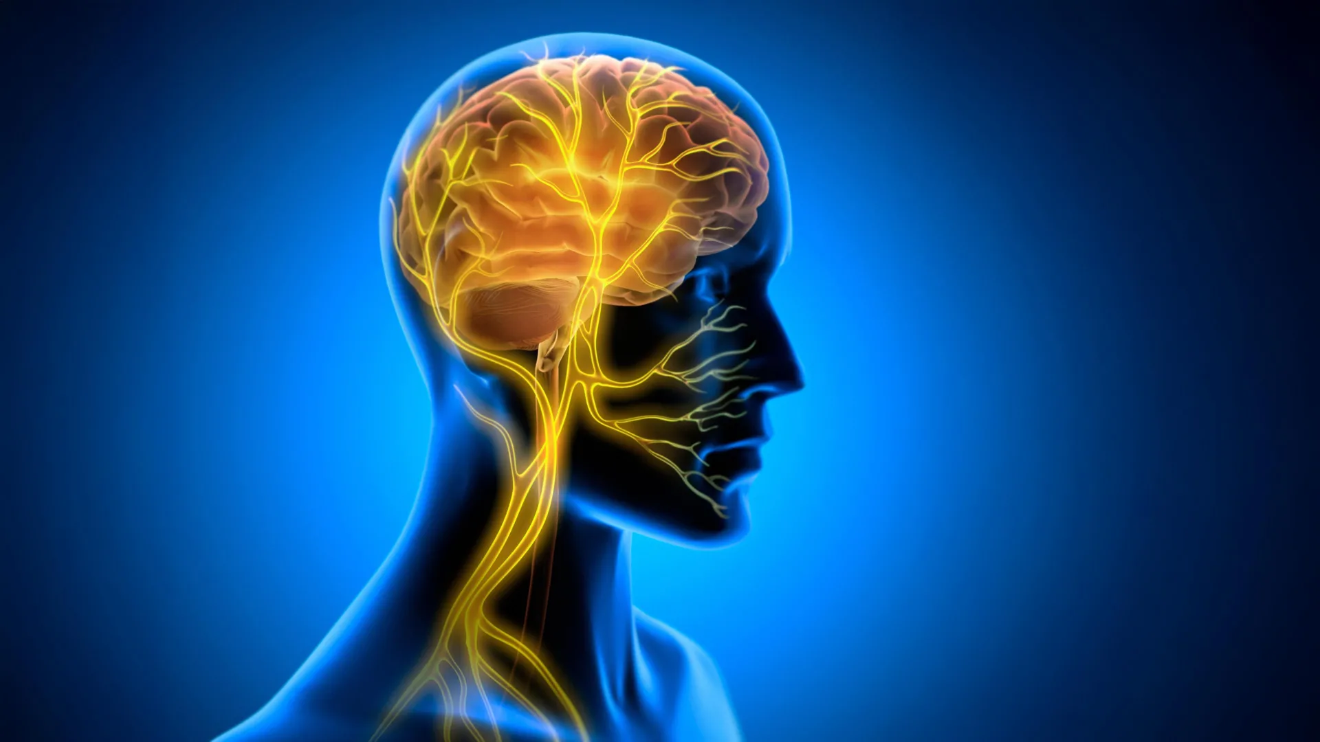
To understand the magnitude of the Tohoku University discovery, one must examine the anatomy and function of the vagus nerve. Originating in the brainstem, the vagus nerve—aptly named from the Latin word for "wandering"—descends through the neck and thorax, branching out to innervate nearly all major internal organs, including the heart, lungs, and gastrointestinal tract. It serves as the primary information superhighway of the autonomic nervous system, facilitating continuous, bi-directional communication between the visceral organs and the central nervous system. Approximately eighty to ninety percent of its fibers are afferent, meaning they continuously transmit sensory data from the body’s internal environment upward to the brain, while the remaining efferent fibers carry regulatory commands downward.
For years, clinical applications of this pathway have focused primarily on vagus nerve stimulation (VNS), a medical intervention that delivers electrical impulses directly to the nerve trunk. VNS is already an FDA-approved therapy for drug-resistant epilepsy and severe, treatment-resistant depression, and it has increasingly been investigated for its utility in stroke rehabilitation and tinnitus management. Historically, neuroscientists believed that VNS exerted its therapeutic and cognitive effects solely through neuromodulation—specifically by prompting the release of key neurotransmitters such as norepinephrine, serotonin, and acetylcholine in brain regions responsible for attention and plasticity.
However, the team at Tohoku University hypothesized that VNS might trigger an additional, fundamental physiological shift: altering the micro-vascular architecture and blood volume dynamics within specific brain regions actively engaged in the learning process. This hypothesis shifted the focus from purely electrical and chemical signaling to the metabolic and hemodynamic environment of the brain itself.
Experimental Design and Methodology
To test this hypothesis, the research team designed a controlled experiment utilizing murine models. They engineered a specialized, miniaturized cuff electrode capable of being permanently yet safely implanted around the left cervical vagus nerve of the mice. This technological refinement allowed for precise, repeatable delivery of electrical stimulation without impeding the subjects’ natural movement or physiological functions.
The behavioral paradigm chosen for the study was the horizontal optokinetic response (HOKR) learning task, a well-established, cerebellum-dependent motor learning model. During the HOKR task, mice were placed in a controlled visual environment surrounded by moving vertical stripes. The task required the animals to reflexively track the moving visual stimuli using precise, involuntary eye movements. This biological mechanism is functionally identical to the optokinetic reflex humans experience when watching passing scenery from the window of a moving train or platform.
Crucially, the researchers structured the timing of the intervention with precise temporal separation. Rather than administering vagus nerve stimulation concurrently with the training tasks—a method frequently explored in prior studies—they delivered the stimulation exclusively after the training sessions concluded. This deliberate delay allowed the researchers to isolate the effects of VNS on memory consolidation, the critical post-learning window during which fragile short-term neural traces are stabilized and integrated into long-term memory networks.
Chronology and Key Behavioral Findings
The timeline of the experiment revealed a clear, statistically significant divergence between control groups and subjects receiving post-training VNS. During the actual training phases, the application of vagus nerve stimulation produced no immediate, observable enhancement in motor performance. The mice receiving stimulation performed identically to their non-stimulated counterparts while tracking the moving visual stripes.
However, a dramatic shift in performance emerged during subsequent testing sessions across the following days. On Days 3 through 5 post-training, the mice that had received the post-training vagus nerve stimulation demonstrated substantially superior long-term retention and motor execution compared to the control group. Their ability to track the moving stripes with precision had stabilized at a significantly higher proficiency level.
This chronological pattern provides compelling evidence that vagus nerve stimulation does not merely boost performance through acute arousal or temporary focus. Instead, it actively engages biological mechanisms that operate during the offline consolidation phase.
"The key point is that VNS was delivered only after training," explains Professor Ko Matsui, senior author of the study. "Our findings suggest that VNS may open a hidden window of opportunity for enhanced learning by making the brain environment more receptive to long-lasting change."
Rhythmic Vascular Dynamics Inside the Cerebellum
To uncover the microscopic changes responsible for this prolonged behavioral enhancement, the Tohoku University team utilized advanced fiber photometry to monitor real-time physiological activity within the cerebellar flocculus—a localized region of the cerebellum specifically responsible for processing the HOKR eye-movement task.
The neuroimaging data revealed a striking physiological response following the delivery of post-training vagus nerve stimulation. A single round of VNS triggered a distinct two-phase vascular reaction within the cerebellar tissue. Initially, local blood volume experienced a brief, rapid decrease, which was subsequently followed by a delayed, sustained increase in blood flow. When the stimulation was administered in repeated cycles, it induced synchronized, rhythmic oscillations in regional blood volume.
Further data analysis established a direct correlation between these vascular rhythms and behavioral learning outcomes. Subjects that exhibited larger, more pronounced blood volume oscillations following VNS demonstrated significantly superior motor learning retention by the fifth day of the experiment. This discovery links the somatic nervous system directly to the cerebrovascular metabolic environment, indicating that rhythmic fluctuations in blood delivery may act as a physical catalyst for synaptic plasticity.
"Our brains may be more strongly influenced by the body than we imagine," states lead author Junyu Chen. "By tuning the brain’s metabolic environment, including rhythmic vascular movements, we may eventually unlock capacities that would otherwise remain latent."
Expert Perspectives and Broader Scientific Analysis
The implications of the Tohoku University study extend far beyond the parameters of murine optokinetic research. Neuromodulation specialists and cognitive neuroscientists outside the immediate research group have noted that the findings offer a unifying framework for understanding how physical health, systemic circulation, and cognitive function are inextricably linked.
Dr. Elena Rostova, a neurovascular researcher at the Institute for Advanced Neurological Studies who was not involved in the study, points out the profound clinical potential of these findings. "For decades, neurorehabilitation has treated the brain as an isolated computational unit that simply needs more cognitive repetition to recover or improve," Dr. Rostova remarks. "This research demonstrates that the brain’s capacity to store new motor patterns is gated by systemic physiological states. By harnessing peripheral pathways like the vagus nerve, we may soon be able to artificially induce optimal neuroplastic windows for patients recovering from strokes, traumatic brain injuries, or neurodegenerative conditions."
Furthermore, sports science and educational institutions are likely to monitor future developments closely. The ability to artificially open "windows of opportunity" for memory consolidation could eventually translate into non-invasive, targeted therapies or protocols designed to accelerate skill acquisition in high-performance athletes, surgeons, musicians, and military personnel.
Implications for Future Research and Therapeutic Development
Despite the promising nature of the findings, the research team emphasizes that significant work remains before these insights can be translated into human clinical applications or consumer technologies. Future phases of the research program will focus on refining stimulation protocols, identifying optimal frequency and duration parameters, and mapping out the precise molecular signaling cascades that connect vagus nerve activation to local cerebrovascular rhythms.
Additionally, scientists must determine whether these two-phase vascular responses and subsequent memory enhancements are localized primarily to the cerebellum or if they represent a universal mechanism applicable across multiple distinct brain regions, including the hippocampus and neocortex, which govern declarative and spatial learning.
As investigations into the brain-body connection continue to accelerate, the boundary between somatic health and cognitive capability becomes increasingly porous. By proving that the body can actively signal the brain to optimize its own metabolic environment for long-term memory retention, the Tohoku University study marks a pivotal step toward a more integrated, whole-body understanding of human learning and neuroplasticity.
