
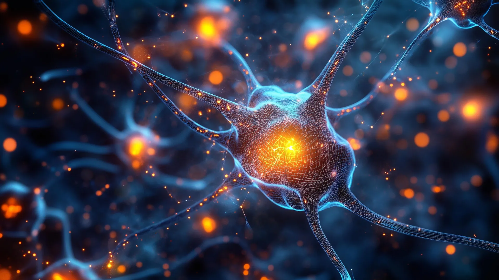
Scientists have identified a group of neurons located in an ancient region of the brain that plays a key role in helping animals focus. These cells appear to improve attention by filtering out distractions and directing the brain toward the most important information. This groundbreaking discovery, made in mice by researchers at Johns Hopkins University, points to a brain system that is shared by all vertebrates, including humans. The findings, published in the prestigious journal Nature Communications and highlighted as an editorial feature, could eventually pave the way for more precise treatments for attention-related disorders such as Attention-Deficit/Hyperactivity Disorder (ADHD) and autism.
Unraveling the Mysteries of Selective Attention
The ability to focus, to selectively attend to relevant stimuli while ignoring distractions, is a fundamental cognitive process essential for survival and interaction with the environment. From navigating a bustling marketplace to deciphering a complex conversation in a noisy café, this capacity, known as selective spatial attention, allows organisms to process a constant stream of sensory information and prioritize what truly matters. Difficulties with this critical function are increasingly recognized as hallmarks of neurodevelopmental conditions like ADHD, characterized by impulsivity and inattentiveness, and autism spectrum disorder, which can involve sensory processing challenges.
For decades, the prevailing scientific understanding largely attributed the intricate mechanisms of attention to the prefrontal cortex, a highly developed region of the brain particularly prominent in humans and other primates. This area is associated with executive functions, planning, and decision-making. However, this explanation presented a significant evolutionary puzzle: how do animals with less developed prefrontal cortices, such as birds and fish, exhibit sophisticated attentional capabilities? This question underscored the need to explore older, more evolutionarily conserved brain structures.
The Brainstem’s Ancient Role in Attentional Filtering
The research team at Johns Hopkins University, led by neuroscientist Shreesh Mysore, embarked on a journey to uncover the ancient neural underpinnings of attention. Their investigation focused on an evolutionarily old region of the brainstem, a fundamental part of the central nervous system that connects the cerebrum and cerebellum to the spinal cord. This area is responsible for many basic life-sustaining functions, including breathing, heart rate, and sleep-wake cycles, but its role in higher cognitive processes like attention was less understood.
"If we really go back in evolution, for hundreds of millions of years, birds have had this ability, fish have had this ability. And they do not typically have a highly developed prefrontal cortex, so how does the brain solve this problem?" explained lead author Ninad Kothari, a postdoctoral fellow in the university’s Department of Psychological and Brain Sciences. "We were able to identify an evolutionarily old region in the brainstem which affords this ability."
The researchers hypothesized that a network of inhibitory neurons within the brainstem might be responsible for this conserved attentional mechanism. These neurons, present across a vast spectrum of vertebrate species, were the subject of earlier investigations by Mysore and his colleagues in birds, frogs, and turtles, hinting at their fundamental importance.
Experimental Design: Mimicking the Human Attentional Challenge
To rigorously test their hypothesis, the scientists designed a sophisticated behavioral experiment for mice that mirrored the challenges faced in human attention studies. The mice were trained to perform a visual task where they were presented with cues on a screen. Their objective was to respond to specific visual information displayed directly in front of them while deliberately ignoring distracting cues that appeared at the periphery of their vision. Successful completion of the task required the mice to actively filter out irrelevant stimuli and focus on the target information, a direct analog of selective spatial attention.
The mice initially performed this task with remarkable proficiency, demonstrating their innate ability to engage in selective attention. This baseline performance served as a critical control for subsequent experimental manipulations.
Disabling the Focus: The Impact of Neuron Inactivation
The pivotal moment in the study came when the researchers employed advanced optogenetic techniques to temporarily switch off the activity of the identified brainstem neurons. This precise genetic manipulation allowed them to selectively silence these specific neuronal populations without affecting other brain functions or causing general distress to the animals.
The immediate consequence of this neuronal inactivation was striking. The mice’s performance on the attention task deteriorated dramatically. They became profoundly "hyper-distractible," as described by Kothari. Even faint, peripheral distractions that they had previously ignored with ease now captured their attention, disrupting their ability to focus on the primary task.
"A hallmark of ADHD is that even faint distractors draw attention away — and that’s exactly what we see here when these neurons are silenced," stated senior author Shreesh Mysore. "But the very next day, when the neurons are turned back on, the same animal can ignore distractors again, even very strong ones."
Ruling Out Alternative Explanations
To ensure that the observed deficits were directly attributable to impaired attention and not to other sensory or motor impairments, the research team conducted a series of additional, carefully controlled experiments. They systematically ruled out the possibility that the mice were failing the task due to vision problems, such as an inability to see the cues, or motor difficulties, such as an inability to make the required responses.
These rigorous investigations confirmed that the animals’ primary deficit was not in their sensory perception or motor execution, but rather in their capacity for cognitive evaluation and selection. The experiments unequivocally demonstrated that the inactivation of the brainstem neurons specifically impaired the mice’s ability to weigh competing pieces of information and direct their attention to the most relevant signal.
"The only thing impaired was their ability to take the competing pieces of information, compare them, and pay attention to the location with the most important information," Mysore elaborated. "This part of the brain is like an attentional selection engine. It helps solve the question: ‘What is most important information I should pay attention to right now?’"
Implications for Human Neurological Conditions
The discovery of this ancient brainstem circuit’s role in attention holds profound implications for understanding and potentially treating attention-related disorders in humans. Given that this brain region and its neuronal populations are conserved across vertebrates, the researchers are optimistic about their potential relevance to human cognition.
"All the evidence to date suggests that these neurons exist in humans too," Mysore affirmed. "But are they responsible for selective spatial attention in humans? An exciting hypothesis is that they play a crucial role."
The findings open up a new avenue of research into the neurobiological basis of conditions like ADHD and autism. Current treatments for ADHD, for example, often involve stimulant medications that affect neurotransmitter systems like dopamine and norepinephrine, indirectly influencing prefrontal cortex function. However, these treatments can have side effects and do not address the root cause for all individuals.
If future studies confirm that these brainstem neurons function differently in individuals with ADHD or autism, it could revolutionize therapeutic approaches. Researchers might be able to develop more targeted interventions, such as pharmacological agents or neuromodulation techniques, specifically designed to fine-tune the activity of these ancient attentional circuits. This could lead to more effective and personalized treatments with fewer side effects.
Future Directions and Broader Impact
The Johns Hopkins research team is now focused on further elucidating the precise mechanisms by which these brainstem neurons influence spatial attention across different vertebrate species. Understanding the intricate neural pathways and signaling cascades involved will be crucial for translating these findings into clinical applications.
Furthermore, the researchers intend to investigate the activity patterns of these neurons in human participants, particularly those diagnosed with ADHD and autism. Advanced neuroimaging techniques and non-invasive brain stimulation methods could provide valuable insights into whether these brainstem circuits are indeed implicated in the attentional deficits observed in these conditions.
The study, which was supported by federal funding, represents a significant leap forward in our understanding of the fundamental neural architecture underlying attention. By shifting the focus from the relatively recent evolutionary development of the prefrontal cortex to the ancient brainstem, the researchers have uncovered a more universal mechanism for attentional control. This paradigm shift not only answers a long-standing evolutionary question but also offers a beacon of hope for developing novel and effective interventions for millions of individuals worldwide affected by attention disorders. The collaborative effort involved researchers Arunima Banerjee, Qingcheng (Jessica) Zhang, and Wen-Kai You from Johns Hopkins University, underscoring the interdisciplinary nature of this significant scientific achievement.
