
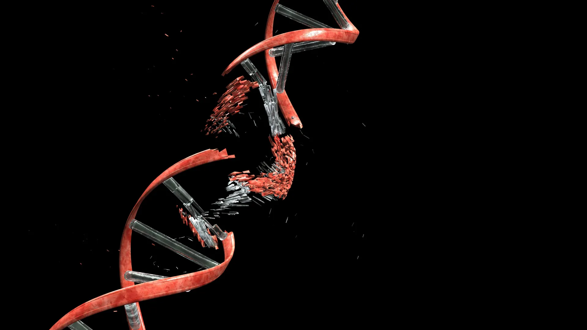
As the intricate architecture of the brain takes shape, a remarkable and previously underappreciated process unfolds: newly formed neurons embark on a perilous journey through densely packed neural tissue to reach their designated locations within the cerebral cortex. This arduous migration, characterized by the need to squeeze through narrow passages between cellular fibers and neighboring cells, was long understood as a feat of cellular navigation. However, a groundbreaking study published in the prestigious journal Nature has unveiled a startling consequence of this developmental odyssey: migrating neurons routinely sustain significant DNA damage, specifically double-strand breaks, a severe form of genomic injury where both strands of the DNA double helix are severed.
This discovery, spearheaded by researchers at Kyoto University’s Institute for Integrated Cell-Material Sciences (WPI-iCeMS) in collaboration with several esteemed institutions, challenges the conventional understanding of DNA damage as solely a harbinger of cellular demise or dysfunction. Instead, the study demonstrates that these double-strand breaks are an intrinsic and surprisingly well-tolerated aspect of healthy brain cortex development. The key to this resilience, the researchers found, lies in the brain’s remarkably efficient DNA repair mechanisms, which swiftly mend the damage before it can precipitate lasting detrimental effects.
"The developing brain appears to have evolved to tolerate and repair the neuronal damage efficiently," stated Professor Mineko Kengaku, the lead author of the study and a distinguished figure at WPI-iCeMS. "But understanding the limits of that tolerance – and what happens when repair is incomplete – brings us closer to understanding a range of neurological conditions." This sentiment underscores the study’s profound implications, shifting the paradigm from viewing DNA damage as an anomaly to recognizing it as a dynamic component of neurodevelopment.
The Mechanical Stress of Migration: Unraveling the Source of DNA Damage
To meticulously investigate the origins of this neuronal DNA damage, the research team embarked on a series of sophisticated experiments designed to replicate the physical hurdles encountered by developing neurons. They ingeniously engineered microchannels, precisely calibrated to mimic the confined and constricted environments characteristic of growing brain tissue. Within these miniature conduits, neurons were guided to navigate the simulated developmental landscape.
Employing advanced fluorescent imaging techniques, the scientists were able to visualize the intricate cellular processes in real-time. Their observations revealed a consistent pattern: as neurons traversed these microchannels, double-strand DNA breaks began to manifest. Crucially, once the cells successfully emerged from the confines of the channels, the observed DNA damage gradually receded. The study reported that the majority of these breaks were repaired within a 24-hour period, allowing the neurons to resume their intended functions without apparent impairment. This observation strongly suggests a dynamic and transient nature to the damage itself.
The study pinpointed a key player in this process: Topoisomerase IIα (Topo IIα), an enzyme that normally plays a critical role in managing DNA topology and relieving torsional stress within the genome. Under normal cellular conditions, Topo IIα functions by transiently cleaving DNA strands to alleviate the strain generated by essential cellular activities, such as replication and transcription, before rejoining them. This mechanism can be conceptually likened to untangling a twisted cable by making a temporary cut, removing the knots, and then seamlessly reconnecting the ends.
However, the researchers elucidated that when neurons are subjected to significant mechanical stress during their arduous migration through tight spaces, this delicate enzymatic process can be disrupted. Topo IIα can become "trapped" mid-cleavage, leaving behind severed DNA strands. The cell then mobilizes its robust repair machinery, primarily the non-homologous end joining (NHEJ) pathway, to re-establish the broken DNA connections. This intricate interplay between mechanical forces, enzymatic activity, and repair pathways forms the core of the study’s findings.
A Unique Neural Resilience: Why Neurons Survive Where Other Cells Falter
A significant aspect of the research involved a comparative analysis of neuronal DNA damage against that observed in other cell types, particularly cancer cells, when subjected to similar microchannel environments. The findings highlighted a striking difference in the cellular response. In cancer cells, DNA damage often occurred in a more random fashion and could readily disrupt vital cellular functions, leading to mutations, further cellular dysfunction, or programmed cell death (apoptosis).
In stark contrast, the DNA breaks observed in migrating neurons exhibited a distinct pattern. The damage was predominantly concentrated in specific regions of the genome that are not actively transcribed or do not harbor essential gene functions. This genomic "hotspot" for damage, coupled with the sparing of critical genes, appears to be the underlying reason for the neurons’ remarkable ability to maintain normal function and viability despite the temporary genomic insult. This specificity suggests an evolutionary adaptation that prioritizes the integrity of crucial genetic information during the critical migratory phase.
When Repair Mechanisms Fall Short: Insights into Neurological Disorders
While healthy developing neurons demonstrate impressive repair capabilities, the study also delved into the consequences of impaired DNA repair. To investigate this, the researchers engineered a model system using mice with a deficiency in Ligase 4, a crucial enzyme within the NHEJ pathway responsible for rejoining double-strand breaks. Specifically, they targeted the newly formed cerebellar neurons of these mice, as the cerebellum is known to be highly sensitive to DNA damage.
Remarkably, these genetically modified mice initially developed without any apparent abnormalities. However, as they progressed into adulthood, subtle yet progressively worsening balance problems began to manifest. These motor deficits closely resemble the symptoms observed in certain human disorders characterized by genome instability, particularly those affecting the cerebellum. This experimental model provides a crucial link between impaired DNA repair during neuronal development and the emergence of neurological symptoms later in life.
The cerebellum, responsible for motor control, coordination, and balance, is densely populated with neurons that undergo significant developmental processes. Disruptions in the precise wiring and function of these neurons, potentially stemming from accumulated unrepaired DNA damage, could explain the observed motor impairments. This finding lends significant weight to the hypothesis that even minor, persistent DNA damage, when left unrepaired, can have profound long-term consequences on brain function.
Broader Implications: Brain Diversity, Disease Etiology, and Future Research
The implications of this research extend far beyond the fundamental understanding of neurodevelopment. The findings strongly suggest that the processes of DNA breakage and repair play a more significant and nuanced role in brain biology than previously appreciated. Researchers are now keen to explore whether these early-life DNA alterations contribute to the inherent diversity observed among individual neurons. Even within the same brain region, subtle variations in DNA repair histories could lead to distinct functional characteristics of individual neurons, influencing synaptic connections and overall network efficiency.
Furthermore, the study opens new avenues for investigating the etiology of neurodevelopmental and neurodegenerative diseases. While the study focused on the early developmental stages, the researchers hypothesize that accumulated DNA damage, or the failure of repair mechanisms over time, could contribute to the onset or progression of conditions such as Alzheimer’s disease, Parkinson’s disease, and various forms of epilepsy. The cerebellum’s role in motor control and its sensitivity to DNA damage also suggest potential links to movement disorders and ataxia.
"It shifts how we think about the neuronal genome," Professor Kengaku emphasized, highlighting the paradigm shift. "All neurons originate from the same DNA, but DNA damage and repair can introduce small genetic differences between individual neurons through a small mechanical journey. Some of that history may be written into the genome itself." This perspective suggests that the very genome of a neuron can bear the imprint of its developmental journey, a concept that could revolutionize our understanding of neuronal identity and plasticity.
The collaborative nature of this groundbreaking research is also noteworthy, involving contributions from the University of Tokyo, the University of Osaka, the National University of Singapore, and the Tokyo Metropolitan Institute of Medical Science, underscoring the global scientific effort to unravel the complexities of brain development and disease. Future research is expected to focus on elucidating the precise genetic and molecular mechanisms that govern the selectivity of DNA damage in neurons, refining our understanding of the specific repair pathways involved, and exploring potential therapeutic interventions aimed at enhancing DNA repair in the context of neurological disorders. The long-term goal is to translate these fundamental discoveries into strategies that can prevent, mitigate, or even reverse the debilitating effects of neurological conditions.
