
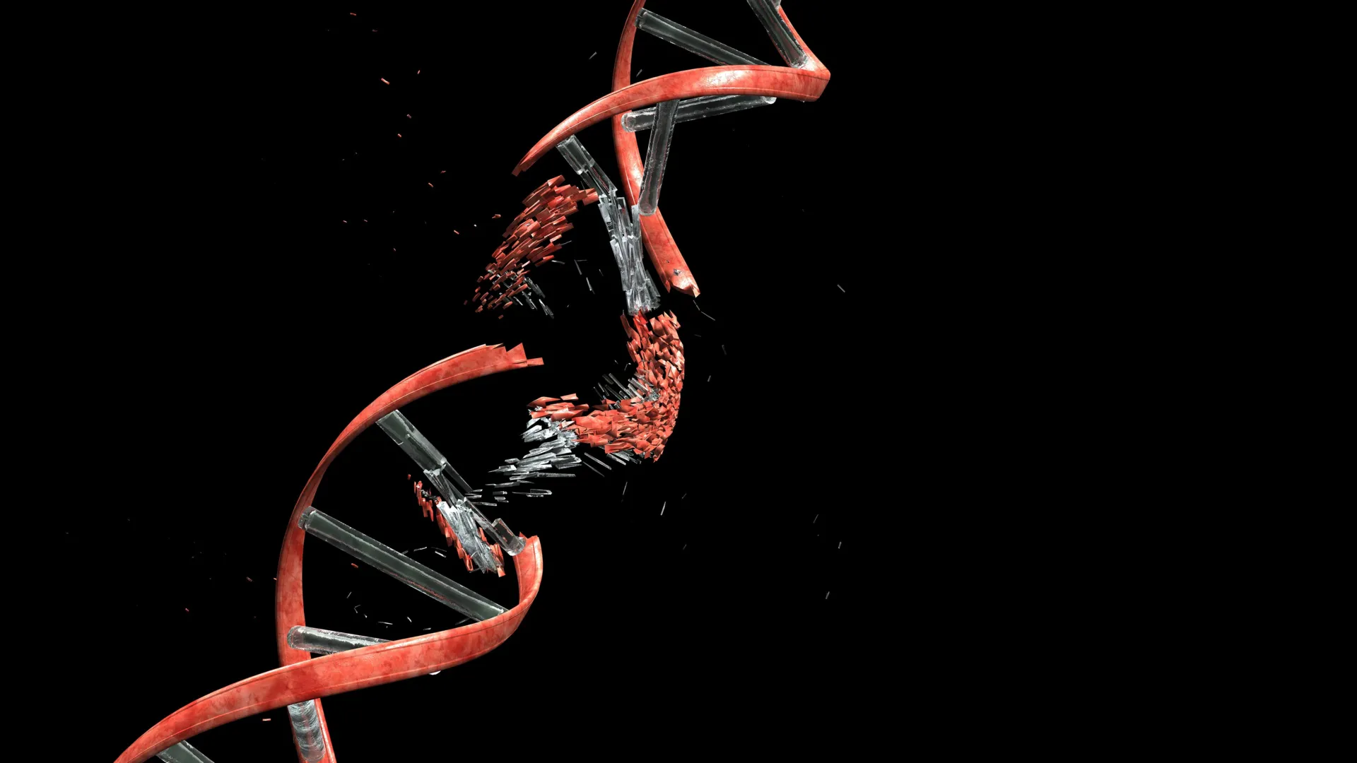
The intricate journey of newly formed neurons through the densely packed cerebral cortex, a critical phase in brain development, has been revealed to involve a surprising level of cellular stress and DNA damage. A groundbreaking study, published in the esteemed scientific journal Nature, has uncovered that these nascent nerve cells routinely sustain double-strand breaks – a severe form of DNA damage – as they navigate the confined spaces and fibrous structures of the developing brain. This unexpected finding challenges previous assumptions about cellular integrity during neurogenesis and highlights the remarkable resilience and sophisticated repair mechanisms inherent in the developing nervous system.
The Perilous Passage of Developing Neurons
The cerebral cortex, the brain’s outer layer responsible for higher-level cognitive functions, is meticulously constructed during embryonic and early postnatal development. This construction process involves the precise migration of billions of neurons from their birthplace in the germinal zones to their designated locations. This migration is not a leisurely stroll; it is a complex and arduous journey. Newly born neurons must traverse a landscape filled with existing cellular structures, glial cells, and a dense network of developing axons and dendrites. The pathways available to them are often narrow, forcing these delicate cells to squeeze through constricting gaps and under significant mechanical pressure.
The study, spearheaded by researchers at Kyoto University’s Institute for Integrated Cell-Material Sciences (WPI-iCeMS) in collaboration with several other leading academic institutions, employed advanced microscopy and genetic engineering techniques to observe this process in unprecedented detail. Their findings indicate that the very act of physical migration through this challenging environment triggers a cascade of events leading to DNA damage.
Unveiling the Double-Strand Break Phenomenon
At the heart of the discovery is the identification of double-strand breaks (DSBs) in the DNA of migrating neurons. DSBs are considered one of the most deleterious forms of DNA damage, as they involve the severance of both strands of the DNA double helix. This type of damage can lead to mutations, chromosomal rearrangements, loss of genetic information, and ultimately, cell death if not accurately repaired. Historically, DSBs have been strongly associated with genotoxic agents like radiation and certain chemicals, and their presence in healthy, developing cells was not widely anticipated.
However, the research team observed that these DSBs were not a sign of cellular demise but rather a transient and, to a degree, expected consequence of neuronal migration. Crucially, the study found that in healthy developing brains, these breaks are remarkably efficient and rapidly repaired. This implies an evolutionary adaptation where the developing nervous system has developed robust systems to cope with and rectify this specific type of damage.
Professor Mineko Kengaku, the lead author of the study and a distinguished researcher at WPI-iCeMS, emphasized the significance of this finding. "The developing brain appears to have evolved to tolerate and repair the neuronal damage efficiently," Professor Kengaku stated. "But understanding the limits of that tolerance—and what happens when repair is incomplete—brings us closer to understanding a range of neurological conditions." This statement underscores the dual importance of the research: illuminating a fundamental aspect of brain development and offering potential insights into neurological disorders.
Mimicking the Microenvironment: The Experimental Approach
To unravel the mechanisms behind this neuronal DNA damage, the researchers meticulously recreated the physical conditions encountered by migrating neurons. They designed and utilized sophisticated microfluidic channels, microscopic conduits engineered to precisely mimic the confined spaces and mechanical stresses that developing neurons face within the brain’s developing tissue. These channels were carefully calibrated to represent varying degrees of cellular density and physical resistance.
Through the use of fluorescently tagged DNA repair markers, the scientists were able to visualize the occurrence and repair of DSBs in real-time as neurons traversed these artificial pathways. The results were striking: as neurons navigated the narrow microchannels, DSBs were observed to form. However, upon emerging from the confined spaces, the damage began to dissipate. The vast majority of these double-strand breaks were successfully repaired within a 24-hour period, allowing the neurons to continue their development and integrate into the neural network without apparent functional impairment. This observation provided compelling evidence that the damage was directly linked to the migratory process and that the cells possessed a potent intrinsic repair capacity.
The Role of Topoisomerase IIβ: A Double-Edged Sword
Further investigation pinpointed the enzyme Topoisomerase IIβ (TopoIIβ) as a key player in the induction of DNA damage during neuronal migration. TopoIIβ is a vital enzyme that plays a crucial role in managing DNA topology, relieving torsional stress that builds up during DNA replication, transcription, and other cellular processes. It achieves this by temporarily creating a break in one or both DNA strands, allowing the DNA to untangle, and then rejoining the broken ends. This process is akin to untwisting a coiled rope to make it manageable.
The researchers discovered that when neurons are subjected to the extreme mechanical forces of squeezing through tight spaces, this crucial enzymatic process can be disrupted. Under such mechanical stress, TopoIIβ can become trapped while the DNA is still broken. This stalled state leaves sections of the DNA helix vulnerable, resulting in persistent double-strand breaks. The cell’s immediate response is to activate its DNA repair machinery, primarily relying on a pathway known as non-homologous end joining (NHEJ), to ligate the severed DNA ends and restore the integrity of the genome.
Neuronal Resilience vs. Other Cell Types
A critical aspect of the study involved comparing the response of migrating neurons to that of other cell types, particularly cancer cells, when subjected to similar microchannel environments. Cancer cells, known for their aggressive proliferation and often aberrant DNA repair pathways, exhibited a different pattern of damage. In cancer cells, DNA damage tended to be more widespread and random, frequently leading to significant cellular dysfunction or triggering programmed cell death (apoptosis).
In stark contrast, the DSBs observed in developing neurons were found to be largely concentrated in specific regions of the genome. These regions were typically those not actively transcribed or involved in the expression of essential genes. This genomic localization is a crucial factor in the neurons’ ability to survive and function. By sparing the critical genes necessary for cell survival and function, the neurons could tolerate the temporary DNA breaks and focus their resources on repair without compromising their fundamental operations. This selective vulnerability and resistance highlight a sophisticated biological mechanism that prioritizes the essential genetic blueprint during development.
When Repair Mechanisms Fall Short: Implications for Neurological Disorders
While the study demonstrated the remarkable repair capabilities of developing neurons, it also explored the consequences when these repair mechanisms are compromised. To investigate this, the researchers created genetically engineered mice whose newly formed cerebellar neurons were deficient in Ligase 4 (LIG4). LIG4 is a critical component of the NHEJ pathway, essential for the efficient repair of double-strand breaks.
Interestingly, these LIG4-deficient mice initially appeared to develop normally, showing no obvious abnormalities in their early stages of life. This suggests that the brain’s developmental processes can compensate to some extent, even with impaired DNA repair. However, as these mice reached adulthood, they began to exhibit progressive, albeit mild, balance problems. These motor deficits are reminiscent of symptoms observed in certain human genetic disorders characterized by genome instability, particularly those affecting the cerebellum, a brain region crucial for coordination and motor control.
This finding provides a crucial link between impaired DNA repair during development and the manifestation of neurological symptoms later in life. It suggests that even subtle, cumulative DNA damage that escapes complete repair can have long-term functional consequences, potentially contributing to neurodevelopmental or neurodegenerative conditions. The cerebellum’s high metabolic rate and complex circuitry may render it particularly susceptible to the effects of accumulated DNA damage over time.
Broader Implications for Brain Health and Disease
The implications of this research extend far beyond the understanding of neuronal migration. It suggests that DNA breakage and repair are not merely incidental events but may play a more fundamental and dynamic role in shaping brain biology than previously appreciated. The study opens up new avenues of inquiry into the origins of neuronal diversity. Professor Kengaku posits, "It shifts how we think about the neuronal genome. All neurons originate from the same DNA, but DNA damage and repair can introduce small genetic differences between individual neurons through a small mechanical journey. Some of that history may be written into the genome itself."
This implies that the unique "history" of each neuron’s migratory journey, imprinted by its encounters with mechanical stress and subsequent DNA repair events, could contribute to subtle genetic variations between individual neurons. These variations, though potentially minor, could influence neuronal function, connectivity, and susceptibility to disease.
Furthermore, the research provides a novel perspective on neurodevelopmental disorders, such as autism spectrum disorder and intellectual disabilities, which are characterized by disruptions in neuronal migration and connectivity. It also offers potential insights into neurodegenerative diseases like Alzheimer’s and Parkinson’s, where cellular stress and DNA damage are known to play contributing roles. Understanding the precise balance between DNA damage and repair in developing neurons could pave the way for new diagnostic markers or therapeutic strategies aimed at protecting neuronal integrity and preventing disease progression.
A Collaborative Endeavor
This significant research was a testament to interdisciplinary collaboration, involving scientists from a consortium of prestigious institutions. The study was conducted through a joint effort involving Kyoto University, the University of Tokyo, the University of Osaka, the National University of Singapore, and the Tokyo Metropolitan Institute of Medical Science. This extensive collaboration brought together diverse expertise in cell biology, genetics, neuroscience, and engineering, enabling the complex experimental design and comprehensive analysis required for such a discovery.
The continued investigation into the intricate relationship between mechanical forces, DNA integrity, and neuronal development promises to deepen our understanding of the brain’s remarkable ability to build and maintain itself, and to shed light on the underlying causes of neurological dysfunction. The finding that the developing brain actively manages and repairs significant DNA damage during its formative stages is a profound testament to the robustness and sophistication of biological processes, even in the face of considerable cellular adversity.
