
Long before a human infant draws its first breath, an intricate and monumental biological orchestration takes place within the womb. Billions of microscopic decisions occur in rapid succession, dictating the eventual architecture of the human brain. At the absolute center of this complex developmental choreography are radial glia—a specialized and remarkably versatile class of neural stem cells. These cells serve as the primary architects of the cerebral cortex, the folded outer layer of the brain responsible for higher-order cognitive functions such as abstract reasoning, long-term memory, complex language acquisition, and sensory processing.
For decades, developmental neurobiologists have recognized that radial glia are central not only to standard human neurogenesis but also to the striking evolutionary expansion of the human cerebral cortex, which dwarfs the cortical surface areas of most other mammalian species. While the vast majority of these specialized stem cells naturally disappear through programmed cell death or differentiation before birth, remnants or reactivated versions of these cells frequently reappear in aggressive forms of brain cancer, a persistent mystery that continues to puzzle oncologists and neuroscientists alike.
Now, two landmark studies published concurrently in the prestigious academic journals Cell and Science have provided an unprecedented, high-resolution look into the precise molecular mechanisms governing how radial glia make their critical developmental choices. Led by researchers at the David Geffen School of Medicine at UCLA, including senior author Dr. Aparna Bhaduri, the research reveals that these master stem cells do not operate on a fixed genetic script alone. Instead, they continuously interpret and respond to two drastically different environmental inputs: internal metabolic signaling and direct physical cues from distant regions of the developing brain. Together, these discoveries bridge long-standing knowledge gaps in human neurobiology and offer profound implications for understanding neurodevelopmental disorders, neuropsychiatric conditions, and oncology.
The Evolutionary Significance and Clinical Paradox of Radial Glia
To understand the magnitude of the recent UCLA findings, one must examine the unique evolutionary trajectory of the human brain. Compared to non-human primates and laboratory rodents, the human cerebral cortex has undergone an extraordinary expansion, characterized by extensive folding (sulci and gyri) that maximizes surface area within the restricted confines of the skull. This expansion underpins humanity’s advanced cognitive capabilities, yet it comes with a vulnerability: the intricate developmental pathways required to build such a complex structure leave wide margins for error.
Dr. Aparna Bhaduri, an assistant professor of biological chemistry at UCLA and a member of both the UCLA Broad Stem Cell Research Center and the UCLA Health Jonsson Comprehensive Cancer Center, has dedicated her career to studying these dynamic cells. "Radial glia are the coolest cells that have ever existed," Bhaduri noted. "They’re really key to making us human. But they’re also at the center of many neurodevelopmental and neuropsychiatric disorders, as well as cancer—so understanding how they make their decisions is one way to start understanding how those conditions arise."
Historically, studying radial glia in living human tissues posed near-insurmountable ethical and technical challenges. Traditional animal models—primarily mice—possess brains that are smooth, significantly smaller, and developmentally distinct from human brains, lacking the specific outer subventricular zone where human radial glia predominantly reside and amplify. Consequently, murine models often fail to replicate human-specific neurodevelopmental disorders or the exact behavioral mechanics of human stem cells. The advent of human pluripotent stem cell technologies, brain organoids, and complex multicellular "assembloids" over the past decade has fundamentally transformed the field, allowing researchers to model human corticogenesis in vitro with remarkable fidelity.
Metabolic Landscapes: Rewriting the Rules of Stem Cell Fate
In the first of the two complementary studies, published in Cell, a multidisciplinary team co-led by Bhaduri’s laboratory and the laboratory of UCLA biochemistry professor Dr. Heather Christofk constructed a high-resolution, comprehensive metabolic atlas of the developing human cerebral cortex. Co-first authors Jessenya Mil and Jose Soto spearheaded the exhaustive biochemical analysis, which combined donated human embryonic and fetal tissue samples with advanced human brain organoid models.
For decades, mainstream neurobiological dogma held that cellular metabolism—the chemical processes by which cells convert nutrients into energy and building blocks—played a strictly supportive, housekeeping role in the background of tissue development. However, the UCLA team’s spatial and biochemical mapping pointed to an entirely unexpected conclusion: metabolism acts as an active driver and regulatory switch capable of directly dictating which cellular lineages neural stem cells choose to produce.
Specifically, the researchers discovered that human radial glia rely heavily upon the pentose phosphate pathway. This vital metabolic route processes glucose molecules to generate the pentose sugars and reducing equivalents required by rapidly dividing cells for nucleic acid and lipid biosynthesis. When the investigative team experimentally manipulated this pathway—either by restricting glucose availability or pharmacologically inhibiting key enzymatic steps within the pentose phosphate cascade—the behavior of the radial glia shifted dramatically. Rather than continuing their standard trajectory, the stem cells altered their production output, prematurely generating increased numbers of inhibitory interneurons and other specialized cell types that typically emerge during later stages of corticogenesis.
"What was surprising is that metabolism isn’t just a passive thing that happens in the background," Dr. Bhaduri explained. "It can really control how stem cells make decisions."
This paradigm-shifting discovery carries significant downstream implications for maternal-fetal medicine. By demonstrating that environmental metabolic states directly alter stem cell fate decisions, the findings provide a mechanistic framework for investigating how maternal nutritional deficits, gestational diabetes, systemic metabolic disorders, and other environmental teratogens can permanently alter fetal brain development. Furthermore, the newly published metabolic atlas serves as an invaluable, publicly accessible reference dataset for global researchers studying bioenergetics in human neurogenesis.
Physical Projections and Early Neural Communication
While the Cell investigation explored the internal metabolic environment of the developing cortex, the second study, published in Science and led by first author Claudia Nguyen, shifted focus to external, physical interactions originating from deep within the subcortex.
Neuroscientists have long established that neurons residing within the thalamus—a deep-seated sensory relay station of the brain—extend long, axonal projections upward toward the developing cerebral cortex to establish complex neural circuits. However, historical anatomical and histological studies of human fetal tissue revealed a peculiar chronological discrepancy: these thalamic wire-like fibers arrive in the cortical plate remarkably early, long before the structural synapses and functional neural networks are fully established and operational.
This temporal mismatch sparked a persistent scientific inquiry: Why do these thalamic projections arrive so far in advance of their functional integration?
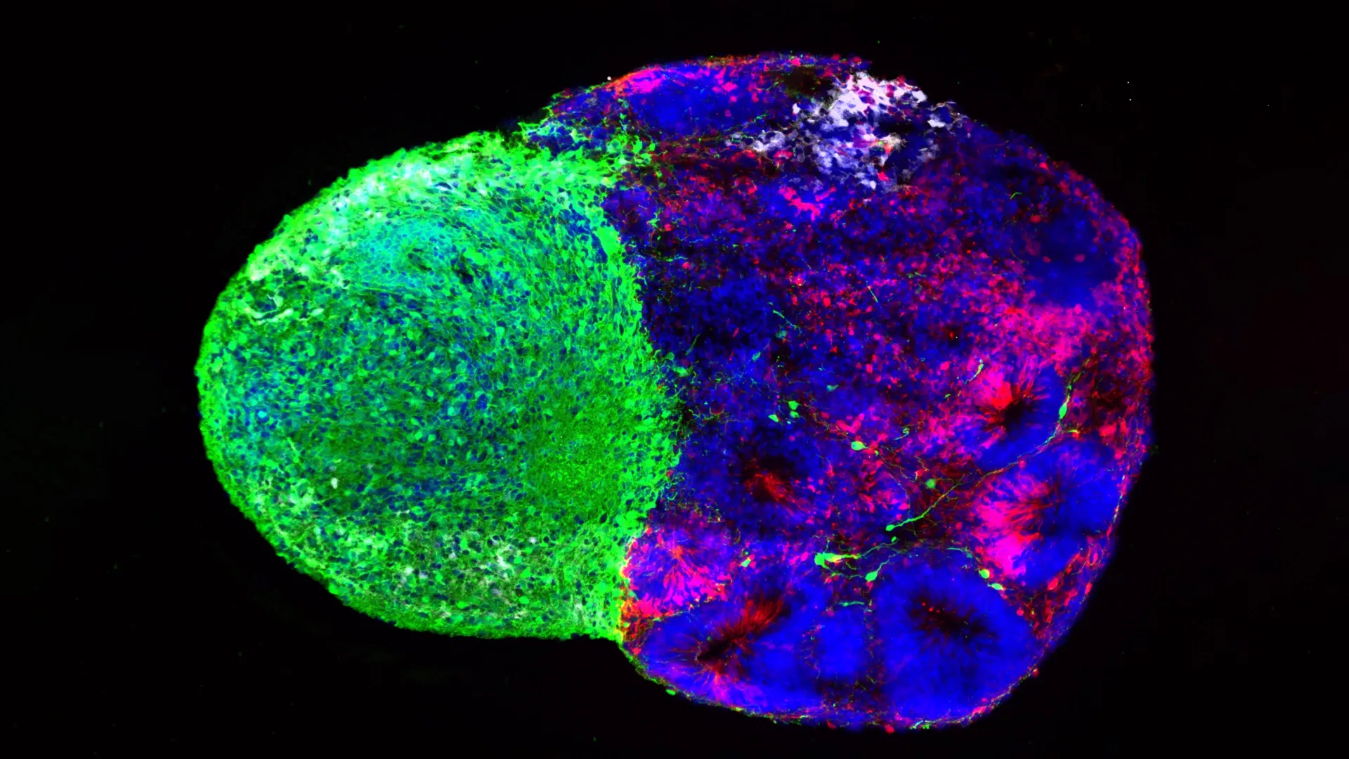
Utilizing sophisticated human stem cell-derived brain "assembloids"—three-dimensional co-culture systems that allow distinct brain regions to grow and interact together in a dish—the UCLA research team uncovered a surprising answer. Rather than merely waiting idly for the cortex to mature, the arriving thalamic projections make direct, physical cell-to-cell contact with radial glia while the cerebral cortex is still actively constructing its layers.
This physical engagement directly alters the developmental programming of the stem cells. Upon physical touch by the thalamic fibers, the radial glia are signaled to increase their production of excitatory glutamatergic neurons—the primary signal-carrying neurons that comprise the bulk of the cerebral cortex. This stimulatory effect was found to be particularly pronounced for upper-layer cortical neurons, a specialized population of cells that have undergone massive evolutionary expansion in the human lineage compared to lower-order mammals.
"We already knew that these projections influence how the cortex develops," Dr. Bhaduri noted. "What we specifically found is that this influence comes through an actual physical connection between the projections and the radial glia—a point of contact that just hasn’t been identified before, and one that very likely does not exist in rodents."
Bridging Developmental Neurobiology and Autism Genetics
To further validate the physiological relevance of this newly discovered physical axis, the UCLA researchers investigated the molecular machinery mediating the contact between thalamic fibers and radial glia. Their investigations led them to NRXN1 (Neurexin-1), a prominent gene widely recognized for its canonical role in helping mature neurons form functional synapses and maintain intercellular adhesion.
Crucially, mutations, deletions, and structural variations within the NRXN1 gene have long been established as high-risk genetic factors strongly associated with autism spectrum disorder (ASD) and other neurodevelopmental conditions characterized by atypical neural connectivity.
To determine whether NRXN1 dysfunction disrupts early stem-cell-level interactions, the team engineered specialized assembloids derived from patient-specific induced pluripotent stem cells carrying known pathogenic NRXN1 mutations. In these mutated laboratory models, the signaling dynamics between the thalamic projections and the radial glia behaved abnormally compared to wild-type control models.
This functional divergence resulted in a distorted balance between the pool of active stem cells and the specific proportions of neurons subsequently generated. By linking a classic synaptic gene to early stem cell behavior, the research offers an innovative window into etiology, suggesting that the developmental roots of certain neurodevelopmental conditions like autism may begin much earlier than previously conceptualized—not merely at the level of mature synaptic transmission, but during the primary construction phases of the cerebral cortex.
Technological Evolution and Future Horizons in Neuroscience
Synthesizing the insights gained from both studies reveals a unified overarching principle: radial glia do not develop in a vacuum. Whether responding to internal metabolic shifts driven by glucose availability or physical tactile cues delivered by long-range axonal fibers, human neural stem cells are in constant, dynamic communication with their surrounding microenvironment.
The success of these parallel investigations underscores the meteoric rise of human brain organoid and assembloid technology. Just ten years ago, researchers lacked scalable, ethically viable experimental systems to interrogate living human neural stem cells directly, forcing an overwhelming reliance on animal models that failed to capture human-specific evolutionary phenotypes. Today, these advanced in vitro platforms allow scientists to ask intricate, mechanistic questions that transcend the limitations of traditional models, bridging the gap between cellular biology and clinical neurology.
Looking forward, Dr. Bhaduri and her collaborators hope these findings will reshape how the broader scientific community conceptualizes brain development, encouraging researchers to treat metabolism and cellular architecture not as static background conditions, but as dynamic, actionable drivers of neurogenesis.
"Ultimately, these studies give us a glimpse under the hood of how these cells make decisions," Bhaduri concluded. "Understanding those decisions is a first step toward understanding normal brain development, disease vulnerability and, potentially, how similar stem-cell programs operate in brain cancer."
This foundational research was made possible through generous financial support and grants provided by the National Institutes of Health, the National Science Foundation, the Brain & Behavior Research Foundation, the Alfred P. Sloan Foundation, the Rose Hills Foundation, the Esther A. & Joseph Klingenstein Fund, the Simons Foundation, the Chan Zuckerberg Initiative, the NIH BRAIN Initiative Cell Atlas Network, the International Foundation for Ethical Research, the UCLA Broad Stem Cell Research Center’s Stem Cell Research Training Program, and the UCLA Health Jonsson Comprehensive Cancer Center and UCLA Broad Stem Cell Research Center Ablon Scholars Program.
