
Researchers at Johns Hopkins Medicine are at the forefront of a promising new avenue for Alzheimer’s disease treatment, thanks to a recently awarded grant from the National Institutes of Health (NIH). This groundbreaking study is delving into the intricate workings of a specific protein in the brain, one that, despite its subtle role, produces a small but significant gas with profound implications for cognitive function and memory. The findings suggest that by understanding and potentially modulating this protein’s activity, scientists may unlock novel therapeutic strategies to combat the devastating effects of neurodegenerative diseases like Alzheimer’s.
Unveiling the Role of Cystathionine γ-lyase (CSE)
At the heart of this research lies the protein known as Cystathionine γ-lyase, or CSE. While perhaps most recognizable for its role in generating hydrogen sulfide (H₂S) – a gas notorious for its pungent "rotten egg" odor – CSE appears to be a critical, albeit previously underappreciated, player in the complex biological processes that underpin memory formation. These pivotal discoveries emerge from meticulously designed experiments conducted on genetically engineered mice, under the leadership of Dr. Bindu Paul, an Associate Professor of Pharmacology, Psychiatry, and Neuroscience at the Johns Hopkins University School of Medicine.
The research, which has been published in the esteemed scientific journal Proceedings of the National Academy of Sciences, is not merely an exploration of a biological curiosity. Its primary objective is to elucidate the precise mechanisms by which CSE functions within the brain. Crucially, the study aims to determine whether enhancing the activity of this protein could offer a protective shield for brain cells, thereby slowing the relentless progression of neurodegenerative conditions, with a particular focus on Alzheimer’s disease.
Hydrogen Sulfide: A Potent Protector of Neurons
Emerging evidence from earlier research had already hinted at the neuroprotective capabilities of hydrogen sulfide in mouse models. These studies indicated that H₂S could play a vital role in safeguarding neurons. However, a significant hurdle in translating these findings into clinical applications has been the inherent toxicity of hydrogen sulfide in higher concentrations. This toxicity renders direct administration to the brain an unsafe proposition. Consequently, the scientific community has been diligently seeking ways to understand how to safely maintain the extremely low, yet vital, levels of H₂S that are naturally present within neurons.
The latest findings from the Johns Hopkins team provide a crucial piece of this puzzle. Their experiments revealed that mice engineered to lack the CSE enzyme exhibited pronounced deficits in memory and learning capabilities. More alarmingly, these CSE-deficient mice displayed increased markers of oxidative stress, significant DNA damage, and a compromised integrity of the blood-brain barrier. These are all pathological hallmarks commonly associated with Alzheimer’s disease, underscoring the critical role of CSE in maintaining brain health. Dr. Paul, who served as the corresponding author for the study, emphasized that these observations provide compelling evidence for CSE’s integral role in preventing neurodegeneration.
A Legacy of Discovery: Building on Decades of Research
The current research stands on the strong foundation of years of pioneering work led by Dr. Solomon Snyder, a Professor Emeritus of Neuroscience, Pharmacology, and Psychiatry at Johns Hopkins. His influential team, in 2014, reported significant findings regarding CSE’s contribution to brain health in mice afflicted with Huntington’s disease. Their investigations utilized mice that were genetically modified to be devoid of the CSE protein. These CSE-deficient mouse models were initially developed in 2008, a pivotal moment when the protein’s association with crucial physiological functions, including blood vessel function and blood pressure regulation, was first established.
Further advancing this line of inquiry, the group’s research in 2021 provided compelling evidence of CSE’s dysfunction in mouse models of Alzheimer’s disease. In a significant breakthrough, they demonstrated that even very small, controlled injections of hydrogen sulfide could offer substantial protection to brain function in these affected mice. While those earlier studies explored the role of CSE in the context of mice carrying additional genetic mutations linked to various neurodegenerative conditions, the most recent research distinctively isolates and examines the specific function of CSE itself.
"This most recent work indicates that CSE alone is a major player in cognitive function and could provide a new avenue for treatment pathways in Alzheimer’s disease," stated Dr. Snyder, a co-corresponding author of the study, who retired from the Johns Hopkins Medicine faculty in 2023. His continued involvement underscores the enduring significance of this research trajectory.
The Direct Link: Memory Loss and CSE Deficiency
To meticulously investigate the intricate relationship between CSE and memory, the researchers conducted a series of comparative analyses. They pitted mice lacking the CSE protein against their normal counterparts, utilizing the same genetically consistent mouse strain that had been developed in 2008. A key experimental paradigm employed was the Barnes maze, a widely recognized test designed to assess spatial memory – the ability of an organism to remember directions and navigate based on environmental cues.
In this particular experimental setup, the mice are tasked with locating a hidden shelter to escape a bright, aversive light. At the two-month mark of their development, both the control group (normal mice) and the CSE-deficient mice demonstrated comparable proficiency, successfully locating the designated shelter within a three-minute timeframe. However, a stark divergence emerged by the six-month mark. The mice lacking CSE struggled significantly to find the escape route, indicating a substantial decline in their spatial memory. In contrast, the normal mice continued to exhibit robust performance, consistently finding the shelter within the expected timeframe.
"The decline in spatial memory indicates a progressive onset of neurodegenerative disease that we can attribute to CSE loss," explained Dr. Suwarna Chakraborty, the first author of the study and a researcher in Dr. Paul’s laboratory. This finding provides a direct, observable link between the absence of CSE and the deterioration of a fundamental cognitive function.
Cellular Corroboration: Brain Changes Mimicking Alzheimer’s Disease
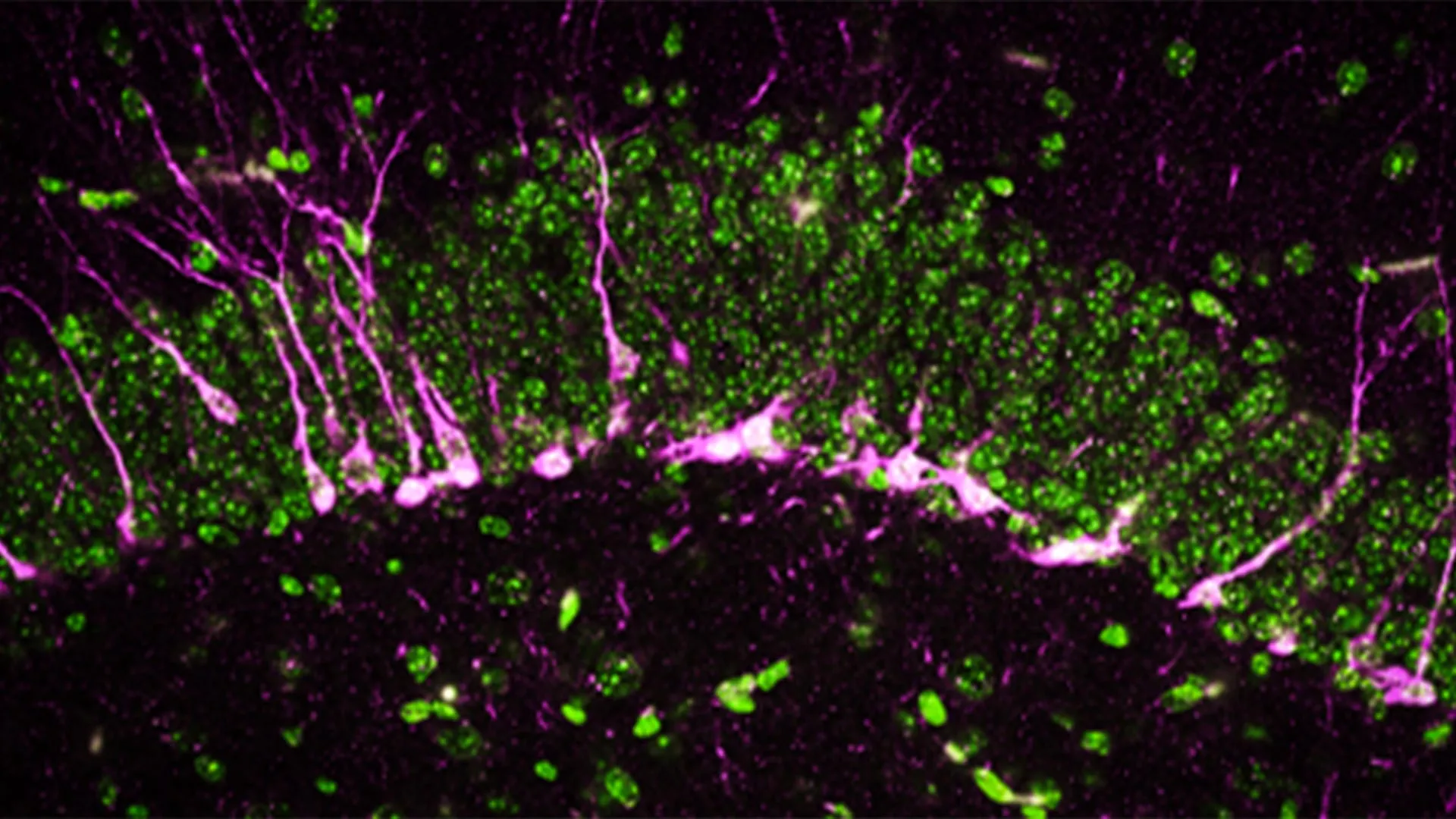
Beyond behavioral observations, the research team also delved into the cellular and molecular consequences of CSE absence within the brain. They meticulously examined how the lack of this protein impacts brain structure and function at a microscopic level. The hippocampus, a region of the brain critically involved in the formation of new memories and learning, relies heavily on the continuous generation of new neurons, a process known as neurogenesis. Disruptions in neurogenesis are a well-established characteristic of various neurodegenerative diseases, including Alzheimer’s.
Employing sophisticated biochemical and analytical methodologies, the researchers observed a significant reduction or complete absence of key proteins essential for neurogenesis in the brains of mice lacking CSE. This cellular deficit suggests a fundamental impairment in the brain’s capacity for self-repair and adaptation.
Further insights were gleaned through the use of high-powered electron microscopes. These advanced imaging tools revealed structural damage within the brains of the CSE-deficient mice. The scientists identified substantial breaks and discontinuities in the blood vessels, a clear indication of damage to the blood-brain barrier. This compromised barrier is another hallmark pathology observed in Alzheimer’s disease, raising concerns about the brain’s ability to protect itself from harmful substances circulating in the bloodstream. Moreover, the study observed that newly formed neurons in these mice encountered difficulties in migrating to the hippocampus, the crucial destination where they would normally contribute to memory consolidation.
"The mice lacking CSE were compromised at multiple levels, which correlated with symptoms that we see in Alzheimer’s disease," stated Dr. Sunil Jamuna Tripathi, a co-first author and researcher in Dr. Paul’s lab. This multi-faceted cellular damage paints a grim picture of the consequences of CSE deficiency, mirroring many of the pathological changes observed in human Alzheimer’s patients.
The Broader Implications: A New Horizon for Alzheimer’s Therapeutics
Alzheimer’s disease represents a formidable public health challenge, affecting over 6 million individuals in the United States alone, according to data from the U.S. Centers for Disease Control and Prevention. The prevalence of this debilitating condition continues to rise, placing an immense burden on patients, families, and healthcare systems worldwide. Despite decades of intensive research, current therapeutic options have proven largely ineffective in consistently halting or even significantly slowing the disease’s inexorable progression.
The findings from Johns Hopkins Medicine offer a beacon of hope. By identifying CSE and its role in hydrogen sulfide production as a potential therapeutic target, researchers are charting a new course for the development of innovative treatments. The strategy would involve devising therapies that can safely modulate CSE activity or enhance the endogenous production of hydrogen sulfide, thereby aiming to protect neuronal integrity, preserve cognitive function, and ultimately slow the devastating march of Alzheimer’s disease.
Funding and Collaboration: A Testament to Scientific Endeavor
This ambitious research initiative has been made possible through substantial financial support from a consortium of prestigious organizations. The National Institutes of Health (NIH) has provided critical funding through multiple grants, including 1R01AG071512, P50 DA044123, 1R21AG073684, O1AGs066707, U01 AG073323, AG077396, NS101967, NS133688, and P01CA236778. Additional support has come from the Department of Defense (HT94252310443), the American Heart Association, the AHA-Allen Initiative in Brain Health and Cognitive Impairment, the Solve ME/CFS Initiative, and the Catalyst Award from Johns Hopkins University. Further contributions have been provided by the Valour Foundation, the Wick Foundation, the Department of Veterans Affairs Merit Award (I01BX005976), the Louis Stokes Cleveland Department of Medical Affairs Veterans Center, the Mary Alice Smith Funds for Neuropsychiatry Research, the Lincoln Neurotherapeutics Research Fund, the Gordon and Evie Safran Neuropsychiatry Fund, and the Leonard Krieger Fund of the Cleveland Foundation.
The collaborative nature of this research is evident in the extensive list of contributors. Beyond Dr. Paul, Dr. Snyder, Dr. Chakraborty, and Dr. Tripathi from Johns Hopkins, the study involved contributions from Richa Tyagi and Benjamin Orsburn at Johns Hopkins; Edwin Vázquez-Rosa, Kalyani Chaubey, Hisashi Fujioka, Emiko Miller, and Andrew Pieper from Case Western University; Thibaut Vignane and Milos Filipovic from the Leibniz Institute for Analytical Sciences in Germany; Sudarshana Sharma from Hollings Cancer Center; Bobby Thomas from Darby Children’s Research Institute and the Medical University of South Carolina; and Zachary Weil and Randy Nelson from the West Virginia University School of Medicine. This interdisciplinary and multi-institutional effort highlights the complexity and the collaborative spirit driving forward crucial advancements in the fight against neurodegenerative diseases. The collective expertise brought to bear on this project underscores its significance and the potential for transformative discoveries.
