McGill University and Douglas Institute Researchers Uncover Distinct Brain Cell Dysfunctions in Depression, Paving the Way for Novel Therapies
Montreal, QC – In a significant stride towards demystifying the biological underpinnings of depression, researchers at McGill University and the Douglas Institute have identified that two specific types of brain cells exhibit differential functioning in individuals diagnosed with major depressive disorder. This groundbreaking discovery, detailed in the esteemed journal Nature Genetics, offers critical insights into the cellular mechanisms of depression and holds immense promise for the development of more targeted and effective therapeutic interventions for a condition that burdens over 264 million people globally and remains a primary cause of disability.
The study, spearheaded by senior author Dr. Gustavo Turecki, a distinguished professor at McGill, a clinician-scientist at the Douglas Institute, and the Canada Research Chair in Major Depressive Disorder and Suicide, marks the first instance where researchers have been able to precisely map gene activity in conjunction with mechanisms that regulate the DNA code to pinpoint specific brain cell types affected by depression. "This is the first time we’ve been able to identify what specific brain cell types are affected in depression by mapping gene activity together with mechanisms that regulate the DNA code," Dr. Turecki stated. "It gives us a much clearer picture of where disruptions are happening, and which cells are involved." This precision fundamentally advances our understanding beyond generalized brain changes to specific cellular culprits.
Leveraging a Unique Resource: The Douglas-Bell Canada Brain Bank
The ability to conduct this nuanced investigation was profoundly dependent on access to a rare and invaluable resource: post-mortem brain tissue samples meticulously collected and preserved at the Douglas-Bell Canada Brain Bank. This specialized collection is globally recognized for its inclusion of donated brain tissue from individuals who lived with psychiatric conditions. Such a comprehensive repository is indispensable for researchers seeking to unravel the intricate biological complexities of mental health disorders at a cellular and molecular level. Without this unique collection, the detailed examination of specific cell types and their genetic profiles would have been exceedingly difficult, if not impossible.
The research team employed state-of-the-art single-cell genomic techniques, a suite of sophisticated methodologies designed to analyze the genetic material of individual cells. By examining RNA and DNA from thousands of these isolated brain cells, scientists were able to discern which cells displayed altered behavior in individuals with depression and to identify specific genetic patterns that might account for these functional divergences. The study’s robust sample size included data from 59 individuals who had received a diagnosis of depression and a control group of 41 individuals without the condition, ensuring a statistically significant basis for their findings. This rigorous methodology allowed for a granular understanding of cellular activity, moving beyond bulk tissue analysis to individual cellular blueprints.
Key Brain Cell Subtypes Show Significant Alterations
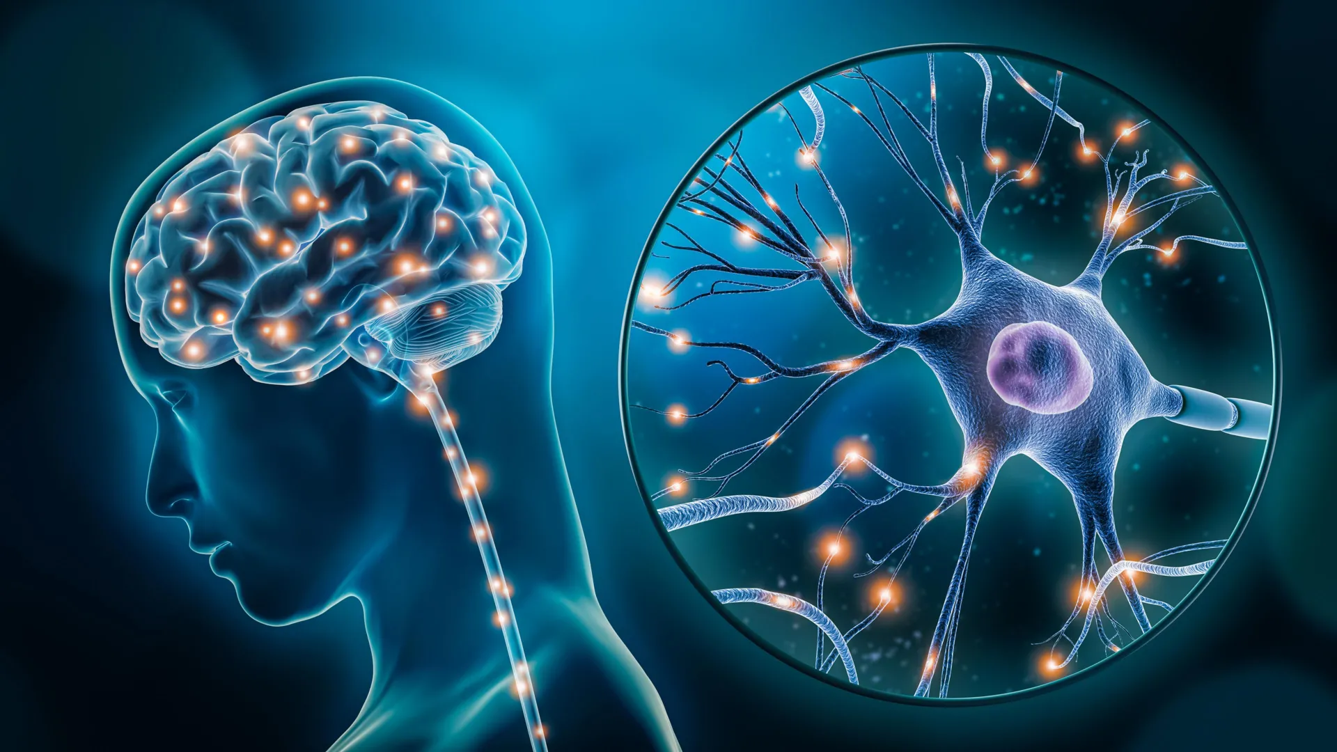
The exhaustive analysis illuminated critical changes in gene activity within two pivotal brain cell populations. The first group comprises excitatory neurons, a fundamental class of neurons responsible for transmitting nerve impulses and playing a crucial role in regulating mood, emotional responses, and the body’s reaction to stress. The second identified cell type was a specific subtype of microglia. Microglia are the resident immune cells of the central nervous system, tasked with a range of functions including clearing cellular debris, responding to pathogens, and importantly, modulating neuroinflammation.
In both of these distinct cell populations – the excitatory neurons and the specific microglial subtype – a substantial number of genes exhibited differential expression levels when comparing individuals with depression to those without. This divergence in gene activity strongly suggests that these vital cellular systems may not be functioning optimally in the context of depression. Such disruptions at the cellular level could provide a compelling biological explanation for the onset and progression of depressive symptoms, moving the understanding of depression from a purely experiential phenomenon to one rooted in measurable biological processes.
Challenging Perceptions: Depression as a Measurable Brain Disorder
This seminal research significantly bolsters the scientific consensus that depression possesses a clear and identifiable biological foundation. It actively challenges historical and lingering misconceptions that have often characterized depression as solely an emotional or psychological affliction, divorced from tangible physiological processes. Dr. Turecki emphasized this point, stating, "This research reinforces what neuroscience has been telling us for years. Depression isn’t just emotional, it reflects real, measurable changes in the brain." This perspective shift is crucial for destigmatizing mental illness and fostering greater acceptance and investment in biological research and evidence-based treatments.
The implications of this research are profound. By pinpointing specific cell types and their associated genetic dysregulations, scientists are no longer working with broad assumptions about brain pathology in depression. Instead, they have concrete targets for investigation. This granular understanding allows for a more precise deconstruction of the complex cascade of events that can lead to a depressive state. It suggests that interventions might one day be tailored not just to managing symptoms but to correcting specific cellular or genetic anomalies.
The Road Ahead: Future Directions in Depression Research
Building upon these foundational discoveries, the research team is now poised to embark on the next critical phase of their investigation. Their immediate focus will be on understanding how these identified cellular differences translate into broader alterations in overall brain function. This will involve exploring the network effects and communication pathways that might be compromised due to the dysregulation in these specific neuronal and microglial populations.
Furthermore, a key objective for the future is to determine whether therapeutic strategies specifically designed to target these affected cell types could yield more effective and potentially curative treatments for depression. This could involve novel pharmacological agents that modulate gene expression in these cells, or perhaps cell-based therapies that aim to restore normal function. The potential for precision medicine in psychiatry, guided by such cellular-level insights, is a tantalizing prospect.
Background and Context: The Evolving Landscape of Depression Research
Depression, classified as Major Depressive Disorder (MDD), is a pervasive and debilitating mental health condition characterized by persistent sadness, loss of interest or pleasure, and a range of emotional and physical problems. Its global prevalence underscores the urgent need for scientific breakthroughs. Historically, treatments for depression have largely relied on psychotherapy and pharmacotherapy, such as Selective Serotonin Reuptake Inhibitors (SSRIs), which target neurotransmitter systems. While these treatments have provided relief for many, a significant portion of individuals experience partial response, treatment resistance, or severe side effects, highlighting the limitations of current approaches and the necessity for novel strategies.
The evolution of neuroscience has progressively shifted the understanding of depression from a purely psychodynamic model to one that acknowledges significant biological contributions. Advances in neuroimaging, genetics, and molecular biology have provided increasing evidence of structural, functional, and chemical alterations in the brains of individuals with depression. However, the precise cellular and molecular mechanisms have remained elusive until recent developments in single-cell technologies. These technologies have enabled researchers to dissect the brain’s complexity at an unprecedented resolution, moving beyond analyzing tissue homogenates to examining the distinct profiles of individual cells within that tissue.
The research conducted by Dr. Turecki and his team represents a crucial milestone in this ongoing scientific journey. By integrating sophisticated genomic analysis with meticulous sample handling and a deep understanding of neuropathology, they have provided a tangible link between the subjective experience of depression and objective biological changes at the cellular level.
Broader Implications and Potential Impact
The implications of this study extend beyond the immediate quest for depression treatments. It reinforces the importance of investing in fundamental biological research for understanding complex human diseases. The methodologies employed, particularly the integration of single-cell genomics and epigenetics, can be applied to the study of a wide array of neurological and psychiatric disorders, potentially accelerating discoveries across the field of mental health.
Moreover, this research has the potential to contribute to a significant destigmatization of mental illness. By demonstrating that depression is associated with measurable biological changes in specific brain cells, it helps to legitimize the condition as a medical disorder, akin to diabetes or heart disease, rather than a personal failing or a sign of weakness. This shift in perception can foster greater empathy, encourage individuals to seek help without shame, and promote increased public and private funding for mental health research and services.
The study also underscores the critical role of biobanks and research infrastructure. The Douglas-Bell Canada Brain Bank, a testament to the foresight and generosity of donors and researchers, has served as the bedrock for this discovery. Such resources are vital for enabling high-impact research that can translate into tangible benefits for patient care.
About the Study and Funding
The findings are detailed in the paper titled "Single-nucleus chromatin accessibility profiling identifies cell types and functional variants contributing to major depression," authored by Anjali Chawla, Gustavo Turecki, and colleagues, and published in the prestigious journal Nature Genetics. This publication signifies the rigorous peer-review process and the scientific community’s recognition of the study’s significance.
The research was made possible through substantial financial support from several key organizations dedicated to advancing health and brain research. Funding was provided by the Canadian Institutes of Health Research (CIHR), Brain Canada Foundation, Fonds de recherche du Québec – Santé (FRQS), and the Healthy Brains, Healthy Lives initiative at McGill University. These collective investments highlight a coordinated effort to tackle complex health challenges through collaborative and innovative scientific inquiry. The sustained support for such foundational research is paramount for continued progress in understanding and treating conditions like depression.
