
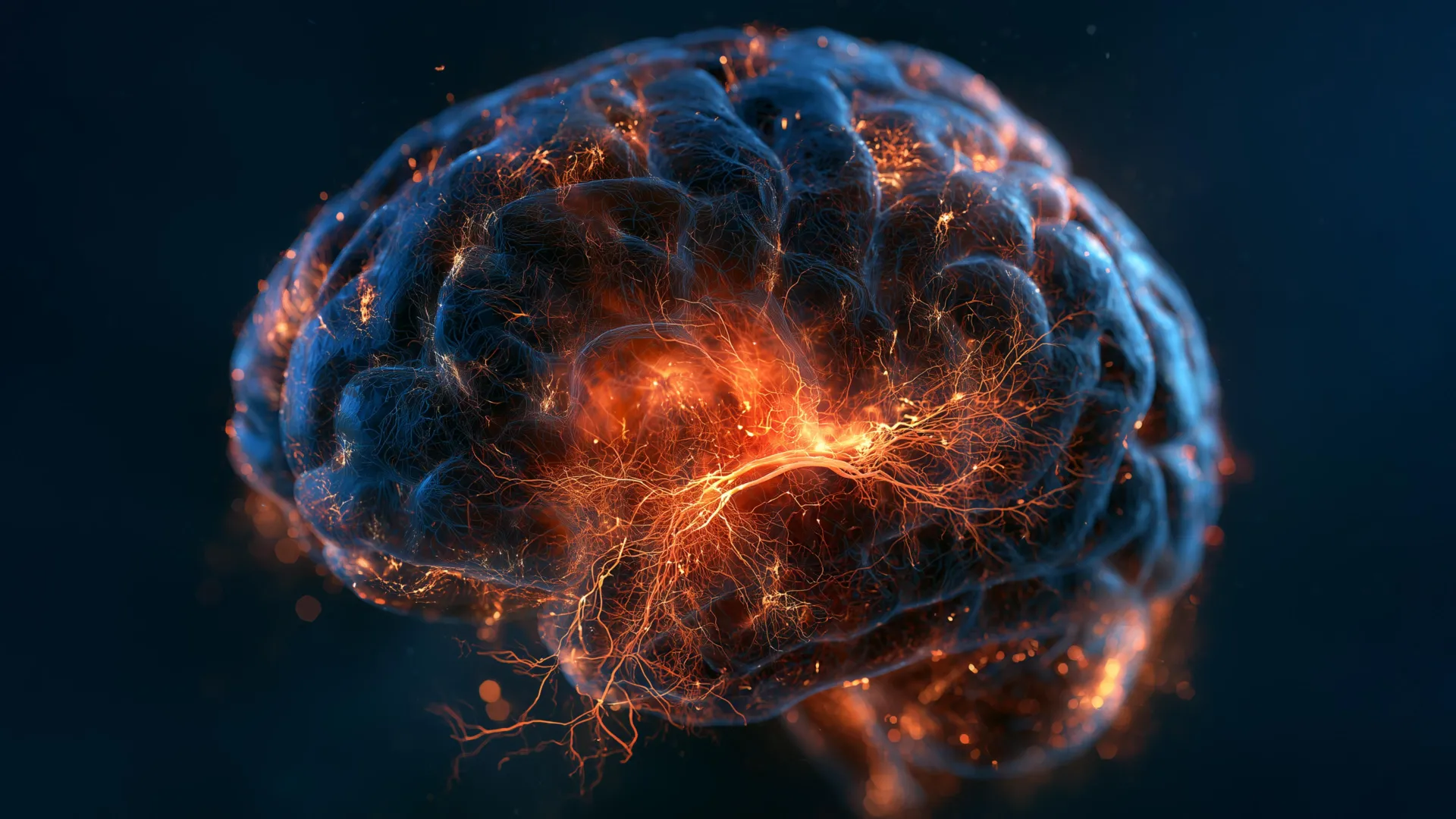
Major depressive disorder has long been conceptualized primarily through the lens of chemical imbalances, most notably involving serotonin and other neurotransmitters. However, a landmark study conducted by researchers at the Columbia University Vagelos College of Physicians and Surgeons and the New York State Psychiatric Institute has upended this traditional framework. Published on August 21, 2026, in the peer-reviewed journal Nature Medicine, the research provides the first direct empirical evidence that neurogenesis—the biological process responsible for generating new neurons—stalls within the adult hippocampus of individuals suffering from major depressive disorder.
The comprehensive study, titled "Dysregulated adult hippocampal neurogenesis in major depressive disorders," was led by Dr. Maura B. Dupont, a professor of psychiatry at Columbia. By analyzing nearly half a million individual brain cells collected post-mortem from donors with depression and healthy control subjects, the research team mapped extensive molecular and cellular disruptions. Their findings suggest that depression is not merely a shortage of chemical messengers, but a systemic failure of the brain’s neurons to adapt to stress, environmental changes, and emotional stimuli. These insights open new pathways for medical science to rethink psychiatric care, potentially paving the way for molecularly targeted therapeutics that could transform how depression is diagnosed and treated globally.
Main Facts and Methodological Breakthroughs
For decades, neuroscientists understood that while the vast majority of the human brain’s approximately 100 billion neurons are generated during embryonic and early childhood development, a small and specialized region known as the hippocampus continues to produce new neurons throughout adulthood. The hippocampus is critically important for processing episodic memories, regulating emotional responses, and navigating spatial orientation.
To investigate the status of this ongoing neurogenesis in clinical depression, Dr. Dupont’s team deployed an array of state-of-the-art single-cell genomic and proteomic techniques. Sequencing operations were executed at the JP Sulzberger Columbia Genome Center, data clustering was managed through Columbia’s Center for Computational Biology and Bioinformatics, and proteomics were analyzed via the Quantitative Proteomics and Metabolomics Center within Columbia’s Department of Biology.
By examining half a million individual cells, the researchers measured the transcriptional activity of every gene within single cells and evaluated whether cellular proteins had undergone pathological alterations. This high-resolution dataset allowed the team to pinpoint the exact spatial localization of affected cells within the hippocampal circuit. The analysis revealed that the disruption in depression extends far beyond halted neurogenesis. The trisynaptic circuit—the primary neural pathway utilized by the hippocampus to establish new emotional memories—exhibited profound signs of inflammation and cellular stress.
Furthermore, researchers identified functional impairments in genes responsible for constructing synaptic connections between neurons, supporting cellular communication, supplying metabolic energy, and facilitating intracellular transport. These widespread anomalies indicate that the structural architecture supporting memory and emotional resilience is profoundly compromised in the brains of individuals with major depressive disorder.
Chronology and Scientific Context
The journey toward understanding the role of adult neurogenesis in psychiatric disorders spans several decades of incremental neuroscientific discovery. Historically, neuroscience dictated that the adult mammalian brain was static, incapable of generating new neurons after early development. However, late 20th-century discoveries challenged this dogma, proving that neurogenesis persists in the subgranular zone of the dentate gyrus within the hippocampus of adult mammals, including humans.
Throughout the 2000s and 2010s, animal model studies—particularly those utilizing murine models—began to link diminished neurogenesis with depressive-like behaviors and demonstrated that successful antidepressant medications could stimulate the growth of newborn neurons. Despite these promising animal studies, obtaining direct, definitive human evidence remained an elusive hurdle due to the technical limitations of studying live human neural tissue and the degradation inherent in post-mortem brain samples.
The Columbia University study represents a critical technological and chronological leap forward. By utilizing advanced single-cell RNA sequencing and high-throughput proteomics on post-mortem human brain tissues, the research team successfully bridged the translational gap between rodent models and human pathology. This breakthrough arrives at a crucial time in psychiatry, where the development of novel pharmacological treatments has stagnated, and clinicians have increasingly sought biological markers to guide personalized medicine.
Supporting Data and the Mechanics of Pattern Separation
To understand the clinical manifestations of halted neurogenesis, researchers focused closely on a cognitive mechanism known as pattern separation. Pattern separation is the brain’s ability to distinguish between similar yet distinct memories and to segregate the emotional weight of past events from current experiences.
In a healthy brain, newborn neurons are uniquely excitable and highly plastic, allowing them to integrate seamlessly into new memory circuits. This plasticity enables the hippocampus to file away distinct experiences into separate neural bins. When neurogenesis stalls, pattern separation becomes impaired. Consequently, memories and their associated emotional connotations blur together.
Dr. Dupont illustrated this phenomenon through clinical observation: "You may be out with a friend for lunch, but she’s tired and doesn’t talk much. With intact pattern separation, you remember this as a unique event. With impaired pattern separation, it becomes mixed with previous memories of feeling rejected, leading you to think, ‘They’re upset with me.’ And I see this a lot in my patients, where they can only retrieve negative information from their memories."
This cognitive bias toward negative memory retrieval contributes significantly to the persistent feelings of worthlessness and hopelessness characteristic of major depressive disorder. Supporting this hypothesis, recent clinical observations involving patients with brain tumors—whose hippocampal neurogenesis was deliberately ablated via localized radiation therapy—exhibited similar cognitive disruptions, reinforcing the causal link between newborn neurons and human emotional regulation.
Genetic Variants, Epigenetic Influences, and Environmental Stress
Beyond cellular counts, the research team analyzed the genetic and epigenetic profiles of the affected brain tissue. The genomic screening revealed altered activity in several specific genes whose genetic variants have historically been linked to an increased risk of major depressive disorder.
Importantly, the study also uncovered widespread epigenetic modifications. Epigenetic mechanisms function similarly to molecular dimmer switches, adjusting the transcriptional activity of genes up or down without altering the underlying DNA sequence. These epigenetic adjustments are highly sensitive to environmental factors, including chronic psychological stress, learning, aging, and chemical exposures.
"These are like dimmer switches that control how active genes are, and they are affected by life experiences such as stress, learning, aging, chemicals, etc.," Dr. Dupont noted.
The immense variability in these genetic and epigenetic alterations offers a compelling biological explanation for a long-standing clinical dilemma: why major depressive disorder manifests with such diverse symptoms from one patient to another. The broad spectrum of molecular disruptions uncovered by the Columbia team suggests that depression is not a uniform monolithic illness, but rather a heterogeneous collection of distinct pathological conditions sharing common clinical symptoms.
Official Responses and Academic Collaborations
The publication of the study in Nature Medicine drew immediate attention from the broader psychiatric and neuroscientific communities, highlighting the collaborative effort required to achieve such granular genomic mapping. The research paper lists an extensive multidisciplinary team of co-authors spanning Columbia University and the New York State Psychiatric Institute, including Madeleine S. Peng, Jialin Jiang, Lucia Polizzi, Tiancheng Shi, Rakshitha Ramkumar, Victor O. Anosike, Giulia Guasoni, Alexandra M. Wamalwa, Madeline B. Mariani, Cheick A. Sissoko, Alexandria N. Tartt, Camille Fulmore, Gorazd B. Rosoklija, Yung-yu Huang, Victoria Arango, Shujuan T. McDonald, Natasha Bitoljanu of Ss. Cyril and Methodius University in Macedonia, Joseph J. Mann, Phi T. Nguyen, Andrew J. Dwork, Lewis M. Brown, René Hen, Hanga Galfalvy, and Maura B. Dupont.
The institutional backing for the project was substantial, drawing on specialized resources across Columbia University Irving Medical Center, the New York State Psychiatric Institute, the JP Sulzberger Columbia Genome Center, and the Department of Biology’s Quantitative Proteomics and Metabolomics Center.
While formal policy responses from global health organizations such as the World Health Organization (WHO) and the National Institute of Mental Health (NIMH) are forthcoming, independent psychiatric researchers have praised the study for establishing a rigorous biological foundation for future translational research. By moving the conversation away from simplistic monoamine deficiency theories, the findings validate decades of clinical observations regarding the profound physical impacts of chronic stress and trauma on the human brain.
Broader Impact and Implications for Future Therapeutics
The ultimate goal of mapping the molecular and cellular architecture of major depressive disorder is to revolutionize clinical treatment paradigms. Dr. Dupont and her colleagues have articulated a visionary objective: to reclassify psychiatric disorders based on their underlying molecular characteristics, mirroring the paradigm shift that has occurred in oncology over the past two decades.
In modern cancer care, tumors are increasingly classified not merely by their anatomical location in the body, but by their specific genetic mutations and cellular features. This precise molecular categorization has enabled the development of targeted immunotherapies and personalized medicine protocols that have dramatically improved survival rates. The research team at Columbia hopes to apply this exact conceptual framework to psychiatry.
"We want to reclassify depression based on its molecular features, similar to what has been done in cancer," Dr. Dupont stated. "Classifying cancers based on their cellular characteristics, not their locations, has led to new and improved treatments. We hope the same will be true for depression and other psychiatric or brain diseases."
If future clinical trials can successfully identify the specific molecular subtypes of depression present in individual patients, physicians may one day bypass the historical trial-and-error approach of prescribing conventional antidepressants. Instead, therapies could be tailored to correct specific cellular deficits—whether by pharmacologically reactivating adult neurogenesis, mitigating localized hippocampal inflammation, or repairing dysfunctional synaptic circuits.
While researchers caution that translating these post-mortem cellular discoveries into safe, effective clinical interventions will require years of rigorous pharmacological development and human clinical trials, the study marks a definitive turning point. By illuminating the microscopic mechanisms that stall neurogenesis and erode neural resilience, modern science has taken a monumental step toward dismantling the biological mysteries of major depressive disorder.
