
The frustration of mastering a new skill—whether it is playing a complex musical passage, executing a precise athletic maneuver, or acquiring a intricate surgical technique—often defies the sheer amount of time and effort invested. While traditional pedagogy has long attributed these disparities in acquisition to innate talent or the repetition of practice hours, emerging neuroscientific research suggests a missing variable: the physiological state of the brain during the consolidation phase. A groundbreaking study conducted by researchers at Tohoku University has demonstrated that the physical condition of the brain, particularly when influenced by signals originating from internal organs, plays a critical role in transforming transient practice into permanent mastery.
Published in the peer-reviewed journal iScience on August 25, 2026, the investigation reveals that stimulating the vagus nerve immediately following training sessions significantly strengthens long-term motor learning in mammalian subjects. This discovery bridges a historical gap in cognitive science by highlighting how somatic communication—signals sent from the body to the brain—actively shapes neuroplasticity. The implications of this research extend far beyond basic murine models, offering a prospective framework for accelerating human skill acquisition and revolutionizing neurological rehabilitation protocols.
The Vagus Nerve as a Bi-Directional Communication Highway
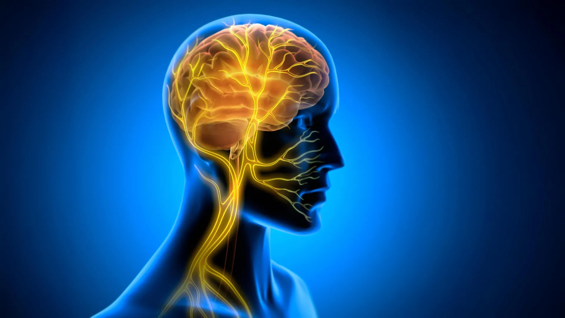
At the center of this discovery is the vagus nerve, one of the longest and most complex cranial nerves in the human and mammalian anatomy. Originating in the brainstem, the vagus nerve extends downward through the neck and thorax, branching out to innervate virtually all major internal organs, including the heart, lungs, and gastrointestinal tract. Functioning as a critical information superhighway, it facilitates continuous, bi-directional communication between the central nervous system and the periphery of the body.
While the brain continuously dispatches regulatory commands downward to modulate heart rate, digestion, and respiratory function, a substantial volume of sensory data travels upward. Approximately eighty percent of the nerve fibers within the vagus nerve are afferent, meaning they carry real-time physiological status updates from the body’s organs directly to the brain. This continuous loop ensures that the central nervous system remains acutely aware of the organism’s internal milieu.
In contemporary medical science, the modulatory capacity of this pathway has been harnessed through vagus nerve stimulation (VNS). Clinically approved by regulatory bodies such as the U.S. Food and Drug Administration (FDA) for decades, VNS therapy has traditionally been deployed to manage drug-resistant epilepsy and treatment-resistant depression. The mechanism typically involves the surgical implantation of a small generator near the clavicle, which sends periodic electrical impulses to the left vagus nerve via attached leads.
Historically, neuroscientists hypothesized that the therapeutic efficacy of VNS stemmed from its ability to prompt the release of vital neurotransmitters—such as norepinephrine, serotonin, and acetylcholine—from subcortical structures like the locus coeruleus. These chemical messengers are known to heighten cortical arousal and foster neuroplasticity. However, the Tohoku University research team suspected that neuromodulation via neurotransmitters might only represent one facet of a much more sophisticated systemic response. They hypothesized that VNS could also instigate hemodynamic and metabolic shifts directly within cerebral microvasculature, creating an optimal physiological environment for memory consolidation.
Experimental Design and Chronology of the Study
To test this hypothesis, the research team at Tohoku University’s Department of Super Network Brain Physiology engineered a specialized experimental framework using murine models. The methodology required precision surgical intervention followed by rigorous behavioral and physiological monitoring over a multi-day timeline.
Chronologically, the investigative process unfolded across several distinct phases:
- Surgical Implantation (Pre-Training Phase): Researchers fitted mice with a custom-designed, miniature cuff electrode. This device was securely yet delicately attached to the left cervical vagus nerve of each subject, allowing for controlled, repeatable electrical stimulation without impeding natural physiological functions.
- Behavioral Training Paradigm: The subjects were subjected to horizontal optokinetic response (HOKR) learning. This well-established, cerebellum-dependent eye movement task trains mice to track moving visual stripes, thereby optimizing their optokinetic reflex. Functionally, this task mirrors the automatic, reflexive stabilization of gaze that humans experience when watching passing scenery from a moving train or platform.
- Post-Training VNS Administration: Diverging from traditional protocols that apply stimulation concurrently with task execution, the researchers administered VNS strictly after each daily training session concluded.
- Longitudinal Assessment (Days 1 through 5): The performance metrics of the mice were meticulously tracked across subsequent days to evaluate the trajectory of skill retention and long-term motor memory consolidation.
- Real-Time Physiological Imaging: Utilizing advanced fiber photometry techniques, the investigative team measured real-time blood volume dynamics near the cerebellar flocculus—a localized brain region directly implicated in the execution and refinement of HOKR learning.
The Temporal Advantage: Why Post-Training Stimulation Matters
One of the most revealing discoveries of the Tohoku University study concerns the timing of the intervention. When VNS was administered, it yielded no immediate, real-time performance enhancement while the mice were actively engaged in the HOKR task. The subjects exhibited standard learning curves during the physical execution of the visual tracking exercise, regardless of whether stimulation was queued.
However, the divergence in performance manifested clearly on subsequent days. Mice that received targeted vagus nerve stimulation immediately following their training sessions displayed significantly superior long-term learning outcomes compared to control cohorts. By the fifth day of the experimental timeline, the stimulated subjects demonstrated markedly enhanced gaze stabilization proficiency.
This delayed-action effect provides critical insight into the neurobiology of learning. Acquisition and consolidation are distinct neurological processes. While acquisition occurs during the active phase of practice, consolidation—the stabilization of a fragile memory trace into a robust, long-term neural network—occurs primarily offline, particularly during post-practice resting states and sleep.
Professor Ko Matsui, a senior investigator on the study, emphasized the strategic importance of this temporal window. "The key point is that VNS was delivered only after training," Professor Matsui noted. "Our findings suggest that VNS may open a hidden window of opportunity for enhanced learning by making the brain environment more receptive to long-lasting change."
This finding challenges the conventional educational and rehabilitative paradigm that assumes optimization must occur synchronously with task practice. Instead, it suggests that manipulating the post-training physiological state can retroactively fortify memory traces, rendering practice sessions exponentially more productive.
Hemodynamic Rhythms and the Brain’s Metabolic Environment
To understand the physical mechanics behind this prolonged learning enhancement, the Tohoku University team peered inside the living brain using fiber photometry. Specifically, they monitored localized blood volume activity within the cerebellar flocculus, the neural hub governing the HOKR task.
The imaging data revealed a precise, two-phase vascular response triggered by a single round of VNS. Immediately following the electrical pulse, local cerebral blood volume experienced a brief, transient decrease. This initial vasoconstrictive dip was systematically followed by a delayed, compensatory surge in blood volume.
Crucially, when the researchers administered repeated rounds of VNS, this cyclical hemodynamic response transformed into sustained, rhythmic oscillations in brain blood volume. These vascular waves were not merely incidental side effects; a direct correlation emerged between the amplitude of these oscillations and the subjects’ behavioral outcomes. Mice that exhibited the most pronounced rhythmic changes in local blood volume during the post-training window ultimately achieved the highest levels of motor learning proficiency by Day 5.
This vascular rhythmicity points to a profound biological reality: the brain’s microcirculatory system is dynamically linked to visceral signals. By modulating the vagus nerve, the researchers effectively tuned the brain’s metabolic environment, optimizing nutrient delivery, cellular waste clearance, and oxygenation precisely when neural circuits were undergoing structural reorganization.
Lead author Junyu Chen elaborated on the broader significance of these physiological dynamics. "Our brains may be more strongly influenced by the body than we imagine," Chen stated. "By tuning the brain’s metabolic environment, including rhythmic vascular movements, we may eventually unlock capacities that would otherwise remain latent."
Implications for Human Rehabilitation, Education, and Medicine
While the study was conducted using murine models, the fundamental physiological architecture of the vagus nerve is highly conserved across mammals, including humans. Consequently, the translational potential of these findings is immense, carrying profound implications across several distinct professional and medical domains.
Neurological Rehabilitation and Stroke Recovery
For patients recovering from stroke, traumatic brain injury, or neurodegenerative conditions, relearned motor skills are often painstakingly slow to acquire. Current physical and occupational therapy regimens rely heavily on sheer repetition. Integrating non-invasive vagus nerve stimulation (nVNS)—which can be administered safely via transcutaneous devices applied to the ear or neck—immediately following physical therapy sessions could dramatically accelerate neural reorganization. By artificially inducing the optimal metabolic and vascular window discovered by the Tohoku team, clinicians might significantly shorten recovery timelines for patients regaining speech, mobility, or fine motor control.
Advanced Skill Acquisition and Training
Beyond clinical applications, the research opens compelling avenues for high-performance fields where rapid, flawless motor learning is paramount. This includes athletic training, surgical residency programs, aviation instruction, and the mastery of complex musical instrumentation. If post-practice somatic interventions can systematically enhance memory consolidation, training methodologies across these industries may evolve to incorporate targeted physiological recovery protocols designed to maximize the neurological return on investment for every hour of physical practice.
Pharmacological and Bioelectronic Medicine
The discovery of a two-phase vascular response driven by peripheral nerve signaling also adds a new dimension to the burgeoning field of bioelectronic medicine. Rather than relying solely on systemic pharmaceuticals—which often produce widespread, undesirable side effects—targeted bioelectronic modulation can induce localized, precisely timed hemodynamic changes within specific cerebellar or cortical sub-regions. This precision could pave the way for a new generation of closed-loop medical devices that monitor a patient’s neurological and cardiovascular state, applying minute vagus nerve adjustments precisely when the brain is poised for structural adaptation.
Future Horizons in Brain-Body Physiology
As the scientific community digests these findings, the research team at Tohoku University is already looking toward the horizon. Future phases of the research will focus on refining stimulation protocols—determining the ideal frequency, duration, and intensity of VNS required to maximize vascular oscillations without inducing habituation or stress.
Furthermore, investigators aim to map out the precise molecular and cellular cascades that link peripheral vagus nerve afferents to microvascular smooth muscle control within the cerebellum and other higher cortical structures. Unraveling the exact biochemical messengers that translate a visceral nerve impulse into a localized cerebral blood wave will provide an unprecedented blueprint of the brain-body axis.
By shifting the focus of neuroplasticity research from isolated cerebral mechanics to the holistic integration of visceral and systemic physiology, this study marks a paradigm shift in our understanding of how humans learn. It affirms that mastery is not merely a product of mental willpower or repetitive exertion, but the result of a finely tuned physiological dialogue between the entire body and the mind.
