
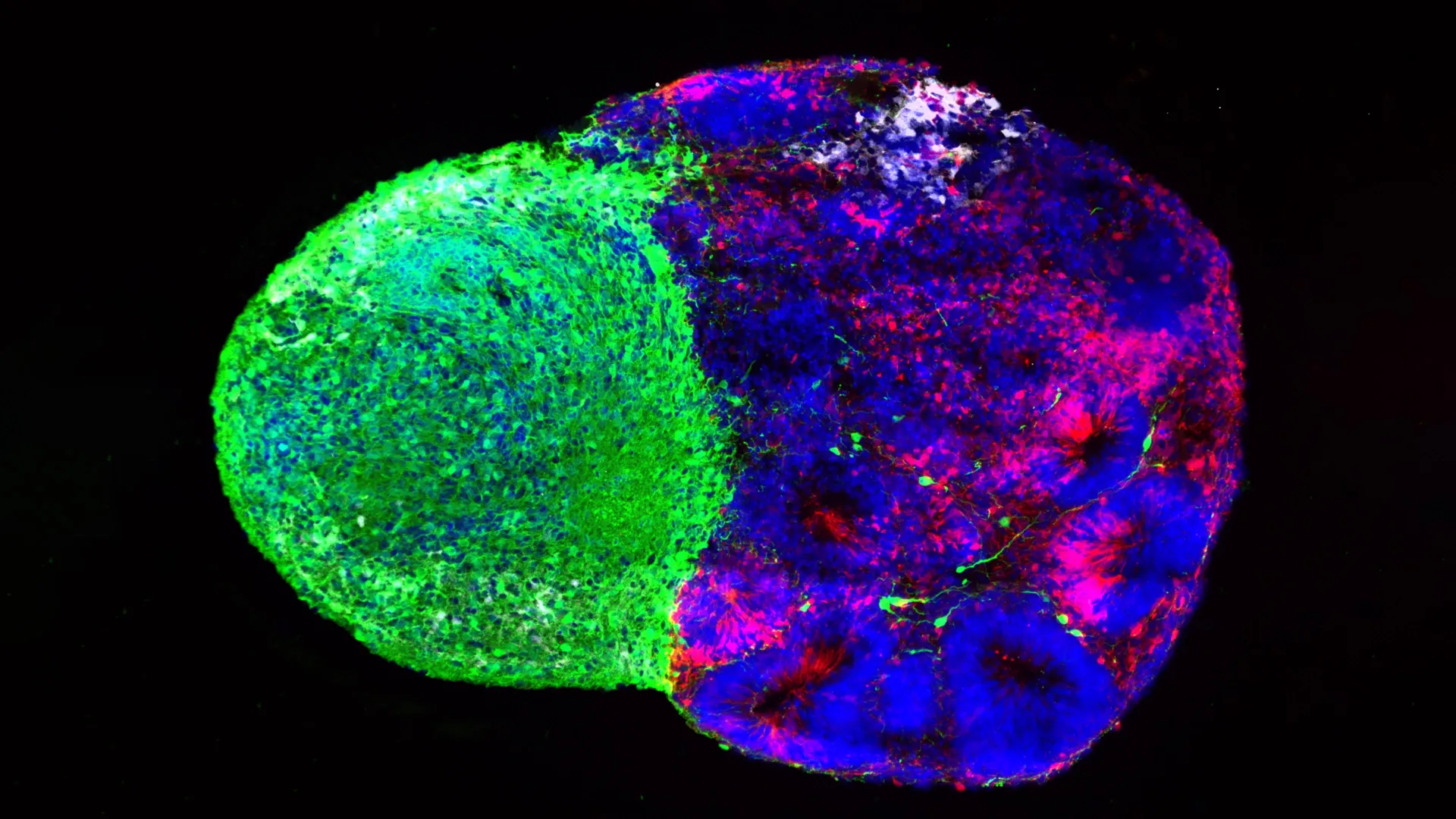
The architectural marvel of the human brain begins long before birth, constructed through an intricate, highly coordinated series of cellular decisions that ultimately define cognition, memory, and language. At the very center of this developmental orchestration are radial glia—specialized neural stem cells that serve as the foundational architects of the cerebral cortex. While neuroscientists have long recognized the vital role of radial glia in generating the expansive human neocortex, the precise molecular and physical cues that dictate their cellular lineage choices have remained largely elusive. Now, two landmark studies published in the journals Cell and Science provide unprecedented resolution into how these master stem cells operate, revealing that metabolic processes and early physical contact from distant brain regions actively drive cortical development. Led by researchers at the University of California, Los Angeles (UCLA), these findings not only illuminate the fundamental mechanics of human neurogenesis but also offer critical new pathways for investigating neurodevelopmental disorders such as autism, as well as certain forms of brain cancer.
The Historical Context of Brain Development Research
To appreciate the significance of the new findings, one must examine the rapid evolution of developmental neurobiology over the past decade. Historically, human brain development was exceptionally difficult to study in real-time. Researchers were forced to rely on post-mortem tissue samples or animal models, primarily rodents, which possess significantly smaller and less complex cortices than humans. While these traditional models yielded fundamental insights into basic cellular biology, they failed to capture the unique features that distinguish human intelligence and cortical expansion.
The landscape shifted dramatically with the advent of human stem cell technology and three-dimensional brain organoids—often referred to as "mini-brains"—alongside more advanced multi-cellular constructs known as assembloids. Developed over the last ten to fifteen years, these laboratory models allow scientists to simulate human embryonic and fetal brain development in vitro. By differentiating pluripotent stem cells into complex tissue architectures, researchers can now observe human-specific developmental pathways that were previously invisible. The UCLA research teams capitalized heavily on these cutting-edge platforms, combining them with donated human fetal tissue to construct detailed atlases of cellular activity and physical interaction during critical windows of prenatal growth.
Metabolic Mapping: How Chemistry Directs Stem Cell Fates
In the first of the two studies, published in Cell, a collaborative team co-led by UCLA biological chemistry assistant professor Aparna Bhaduri and professor Heather Christofk—with co-first authors Jessenya Mil and Jose Soto—ventured beyond traditional genetic signaling to investigate cellular metabolism. By analyzing donated human tissue alongside stem cell-derived brain organoids, the researchers constructed a comprehensive metabolic atlas of the developing human cortex.
The prevailing assumption in developmental biology had long been that cellular metabolism acts merely as a passive engine, providing the baseline energy required for cells to grow and divide. However, the UCLA team’s findings challenge this paradigm, demonstrating that metabolic pathways actively instruct radial glia regarding which types of cells to produce.
Specifically, the researchers discovered that radial glia rely heavily on the pentose phosphate pathway—a metabolic process that utilizes glucose to manufacture the essential molecular building blocks required by rapidly dividing cells. When the research team experimentally restricted the availability of glucose or disrupted the pentose phosphate pathway within the organoid models, the behavior of the stem cells shifted markedly. Rather than producing their standard cellular output, the radial glia began generating higher proportions of inhibitory neurons and other specialized cell types that typically emerge much later in the developmental timeline.
This active participation of metabolism in cell-fate decisions provides a novel framework for understanding how environmental factors during pregnancy—such as maternal nutrition, gestational diabetes, and other metabolic disorders—might alter fetal brain development. Furthermore, the newly published metabolic atlas serves as an invaluable reference tool for the scientific community, charting biochemical shifts across developmental stages with unprecedented precision.
Early Thalamic Projections and Physical Cellular Communication
While the Cell study explored biochemical and metabolic influences, the second study, published in Science and led by first author Claudia Nguyen, examined a radically different source of developmental instruction: structural and physical signaling from deep within the brain.
For years, neuroscientists have documented that neurons residing in the thalamus—a deep-seated relay station that processes sensory and motor signals—extend long, wire-like axonal projections toward the developing cortex. Anatomical studies in humans have consistently revealed a striking chronological anomaly: these thalamic projections arrive at the cortex long before final synaptic connections are established. For decades, the physiological purpose of these early-arriving fibers remained an open question.
Using advanced human stem cell-derived brain assembloids, the UCLA team discovered that these thalamic projections do more than simply wait for target neurons to mature. Instead, the fibers physically make direct contact with radial glia while the cortex is still actively forming.
This physical interaction fundamentally alters the trajectory of the stem cells, prompting them to generate an increased yield of excitatory neurons—the primary signal-transmitting cells of the cerebral cortex. Crucially, this effect was observed to be particularly pronounced for upper-layer cortical neurons, a population of cells that has undergone massive evolutionary expansion in the human brain compared to other mammals. According to researchers, this direct cell-to-cell mechanical communication represents a novel developmental mechanism that likely does not exist in rodents, underscoring the importance of human-specific experimental models.
Implications for Neurodevelopmental Disorders and Genetics
The discovery of direct physical communication between thalamic fibers and radial glia took on heightened clinical relevance when the researchers investigated the molecular machinery mediating the contact. The team identified NRXN1—a gene widely recognized for its role in helping neurons form stable connections—as a key player in the interaction. Mutations in the NRXN1 gene have long been established as significant risk factors for autism spectrum disorder (ASD) and other neurodevelopmental conditions.
To test the functional impact of these genetic anomalies, the researchers engineered assembloids using patient-derived stem cells carrying an NRXN1 mutation. In these mutated models, the signaling dynamics between the thalamic projections and the radial glia were measurably altered. This disruption cascaded through the system, shifting the delicate balance between the maintenance of the stem cell pool and the subsequent generation of neurons.
By bridging the gap between early structural contact, gene expression, and neurodevelopmental pathology, the research offers a plausible mechanistic explanation for how genetic variants associated with autism can manifest as structural anomalies in the cortex long before birth. It suggests that neurodevelopmental conditions may originate much earlier in gestation than previously understood, rooted in the foundational decisions made by embryonic stem cells.
Broader Impacts and Future Directions for Neuroscience
Taken together, these complementary studies present a unified conceptual shift in how neurobiologists view the developing brain. Whether through metabolic programming or physical cellular contact, radial glia do not operate in a vacuum. Their developmental choices are continuously sculpted by a dynamic dialogue with their local and distant microenvironments.
Beyond their implications for normal neurodevelopment and autism, these findings carry significant weight for oncology. During early human development, radial glia proliferate rapidly before most of them disappear prior to birth. However, similar stem-cell programs frequently reactivate later in life during the formation of aggressive brain cancers, such as glioblastoma. By decoding the decision-making logic of radial glia during embryogenesis, researchers hope to gain critical insights into the vulnerabilities of cancer cells that hijack these ancient developmental pathways.
As organoid and assembloid technologies continue to mature, the scientific community anticipates even deeper revelations regarding the unique trajectory of the human brain. While further research is required to translate these laboratory models into clinical interventions, the work led by Bhaduri, Christofk, and their colleagues establishes a robust foundation. By peering beneath the hood of human cellular development, science moves one step closer to untangling the complex origins of cognition, neurological disease, and the biological essence of what makes the human brain truly unique.
Funding and Institutional Support
This extensive body of research was made possible through financial backing and fellowships from a broad coalition of public and private institutions. Primary support was provided by the National Institutes of Health (NIH) and the National Science Foundation (NSF). Additional funding and programmatic support came from the Brain & Behavior Research Foundation, the Alfred P. Sloan Foundation, the Rose Hills Foundation, the Esther A. & Joseph Klingenstein Fund, the Simons Foundation, the Chan Zuckerberg Initiative, the NIH BRAIN Initiative Cell Atlas Network, and the International Foundation for Ethical Research. Furthermore, support was extended via the UCLA Broad Stem Cell Research Center’s Stem Cell Research Training Program, the UCLA Health Jonsson Comprehensive Cancer Center, and the UCLA Broad Stem Cell Research Center Ablon Scholars Program.
