
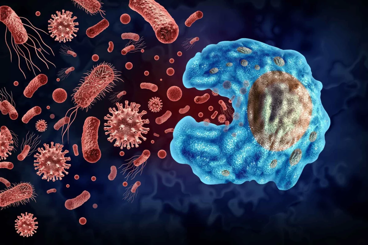
For years, a growing body of scientific literature has suggested that tumors throughout the human body harbor their own unique communities of resident microorganisms. These so-called tumor microbiotas were theorized to play a profound role in shaping cancer biology, influencing everything from tumor progression and immune system evasion to therapeutic response and patient prognosis. However, a landmark study recently published in the journal Cell radically challenges these assumptions, demonstrating that a vast majority of the microbial signals detected within tumor samples are not native inhabitants at all, but rather the result of environmental and laboratory contamination.
The new research, led by scientists at the prestigious Dana-Farber Cancer Institute in Boston, Massachusetts, provides a rigorous reassessment of microbial presence across thousands of cancer genomes. The findings indicate that true, biologically relevant tumor microbiotas are largely restricted to cancers originating in the mouth and the digestive tract. Furthermore, the composition and density of these genuine microbial populations appear to be directly linked to the mutational burden of the cancer cells themselves, offering a fresh perspective on how microbes and malignancies interact.
This comprehensive investigation arrives at a critical juncture in oncology and microbiome research. Over the past two decades, advancements in high-throughput sequencing technologies have allowed researchers to peer deeper into the biological ecosystems of diseases than ever before. Yet, this technological leap has also brought unprecedented challenges, particularly regarding the sensitivity of sequencing tools and their vulnerability to background noise. By rigorously filtering out artifacts and establishing stringent analytical parameters, the Dana-Farber team has introduced a methodological benchmark that is expected to reshape how future cancer microbiome studies are conducted and interpreted.
Background Context: The Complex History of Cancer and Microbes
The intersection of microbiology and oncology is not entirely new; historical precedent firmly establishes that certain microorganisms are capable of driving specific malignancies. For decades, oncologists and microbiologists have recognized definitive causal links between particular infections and specific cancer types. Among the most notable examples is Human Papillomavirus (HPV), which is intricately tied to the development of cervical and certain types of oral cancers. Similarly, the Epstein-Barr virus (EBV) has long been implicated in the pathogenesis of specific gastric cancers and nasopharyngeal carcinomas.
In the realm of bacterial pathogens, Helicobacter pylori stands out as a primary driver of chronic gastric inflammation, peptic ulcer disease, and ultimately, stomach cancer. More recently, extensive research has highlighted the enrichment of specific bacterial species—such as Fusobacterium nucleatum—within colorectal tumors, suggesting that the gut microbiome plays an active role in modulating colorectal carcinogenesis.
Buoyed by these discoveries, researchers began expanding their search to determine whether microbes resided in virtually all human tissues, including tumors outside the gastrointestinal tract. Studies over the past ten years frequently claimed to have identified distinctive microbial signatures in tumors as diverse as breast cancer, brain glioblastomas, lung carcinomas, and prostate malignancies.
However, these findings were frequently met with skepticism by a subset of the scientific community. Critics pointed out that low-biomass samples—tissues containing very small amounts of microbial DNA—are exceptionally prone to contamination during sample collection, DNA extraction, and sequencing library preparation. Reagents, laboratory water, extraction kits, and even ambient laboratory air often carry trace amounts of microbial DNA. When high-throughput sequencers amplify these minute signals, they can easily be misattributed to the biological sample itself, creating the illusion of a tumor-specific microbiome where none actually exists.
Developing a Rigorous Computational Pipeline
To resolve the intense debate surrounding the existence of pan-cancer microbiotas, a research team led by Anders Dohlman at the Dana-Farber Cancer Institute set out to create a failsafe analytical framework. The objective was to design a computational pipeline capable of distinguishing true biological signals from the pervasive background noise of laboratory and environmental contamination.
The newly developed pipeline was engineered to execute several critical functions with high precision. First, it performs an exhaustive depletion of human DNA sequences from genomic datasets, ensuring that host genetic material does not obscure microbial findings. Next, it identifies any remaining microbial DNA sequences and maps them against comprehensive reference databases. Finally, it applies quantitative algorithms to estimate the precise abundance and concentration of microbial material present within the sample.
Before deploying the tool on large-scale clinical databases, the researchers subjected the pipeline to rigorous validation tests. They utilized carefully controlled artificial mixtures containing known proportions of human and microbial DNA. The testing revealed that the pipeline possessed exceptional sensitivity, successfully detecting microbial DNA even when it was present at extremely low concentrations. This high degree of sensitivity was vital to ensure that the tool would not overlook genuine, low-abundance microbes while still maintaining strict specificity against contaminants.
Analyzing More Than 16,000 Tumor Genomes
Armed with this validated computational pipeline, Dohlman and his colleagues conducted a massive retrospective analysis encompassing more than 16,000 tumor genomes spanning a wide spectrum of cancer types. The dataset provided an unprecedented opportunity to map microbial distribution across human malignancies on a global scale.
As expected, the results confirmed that colorectal tumors harbored significantly stronger and more diverse microbial signals than tumors derived from typically sterile environments, such as the brain and the circulatory system. However, a closer examination of the blood and brain tumor samples yielded a telling discovery: despite these tissues being traditionally viewed as sterile, the sequencing data still revealed the presence of low-level microbes. Crucially, these microbes closely matched known environmental and laboratory contaminants, reinforcing the researchers’ hypothesis that many previously reported pan-cancer microbial findings were artifacts of the experimental process.
When the team expanded their analysis across the entire cohort of 16,000 genomes, a striking pattern emerged. The vast majority of cancer types failed to show any convincing evidence of a resident tumor microbiota. The exceptions were strictly confined to cancers originating in anatomical sites directly exposed to the external environment: the mouth, throat, esophagus, stomach, and colorectal tract. In these specific locations, tumors frequently hosted complex and diverse microbial ecosystems comprising various species of bacteria, fungi, viruses, and archaea.
Tumor Mutation Burden as a Key Predictor of Colonization
One of the most provocative findings of the Dana-Farber study relates to why certain tumors host microbes while others remain sterile. Rather than finding that microbial colonization was dictated strictly by the specific histological subtype of the cancer, the researchers discovered that the tumor mutation burden (TMB) was a far stronger predictor.
Tumor mutation burden refers to the total number of genetic mutations found within the DNA of cancer cells. The team observed a direct correlation: tumors carrying a higher burden of DNA mutations tended to contain significantly greater amounts of microbial DNA.
The researchers propose a compelling biological mechanism for this phenomenon. Tumors with high mutation burdens often exhibit altered microenvironments, including distinct metabolic landscapes and modified immune surveillance systems. These localized alterations can create a more permissive environment that allows circulating or locally invading microbes to successfully colonize the neoplastic tissue. Essentially, highly mutated tumors may act as biological sanctuaries where microbes can evade host immune responses more effectively than in normal, healthy tissue or in genetically stable tumors.
Addressing Persistent Uncertainty in Oncology
The publication of these findings in Cell represents a major course correction for the field of cancer microbiome research. For years, conflicting reports have populated scientific literature, leaving oncologists, microbiologists, and patients uncertain about the true clinical relevance of tumor-associated microbes.
While the authors of the study acknowledge that their methodology may have occasionally missed extremely rare or highly fastidious microbes—or potentially misclassified certain ambiguous sequences—they maintain that their overarching conclusions provide much-needed clarity.
"Our analysis helps resolve persistent uncertainty stemming from conflicting reports about the scope and distribution of microbial colonization across cancer sites," the authors noted in their published findings. By establishing a high standard for contamination control, the research effectively draws a line under years of speculative claims regarding widespread pan-cancer microbiotas.
Implications for Future Cancer Research and Diagnostics
The implications of this study extend far beyond theoretical microbiology, carrying immediate consequences for translational oncology, diagnostic development, and therapeutic innovation.
First and foremost, the findings necessitate a re-evaluation of past and future studies that attempt to utilize tumor microbiotas as diagnostic biomarkers or prognostic indicators. If a large proportion of previously reported microbial signals are indeed laboratory or environmental contaminants, any diagnostic assays built upon those findings run the risk of yielding false positives or leading researchers down unproductive clinical pathways. Consequently, research teams worldwide will likely adopt pipelines similar to the one developed by the Dana-Farber institute to re-analyze existing datasets and validate future discoveries.
Second, the confirmation that true microbiotas are localized to cancers of the mouth and digestive tract reinforces the importance of studying host-microbe interactions in anatomically relevant contexts. In colorectal cancer, for instance, understanding how specific bacterial communities interact with mutated cancer cells and influence therapeutic efficacy—such as response to immunotherapy or chemotherapy—remains a vibrant and clinically vital area of study. Researchers can now direct their resources more efficiently toward these biologically validated areas, avoiding the pursuit of phantom microbiotas in sterile tissues like the brain or bone marrow.
Finally, the link established between tumor mutation burden and microbial colonization opens up new avenues for investigating the tumor microenvironment. Understanding how hyper-mutated cancers facilitate microbial survival could yield insights into immune evasion mechanisms, potentially inspiring novel therapeutic strategies that target both the cancer cells and their microbial accomplices.
As the scientific community digests the implications of this comprehensive genomic analysis, the overarching message for the field of oncology is one of rigorous self-correction and methodological refinement. By separating genuine biological phenomena from the persistent shadow of laboratory contamination, researchers are now better equipped to chart an accurate course toward understanding the complex, multifaceted relationships between microbes and human cancer.
