
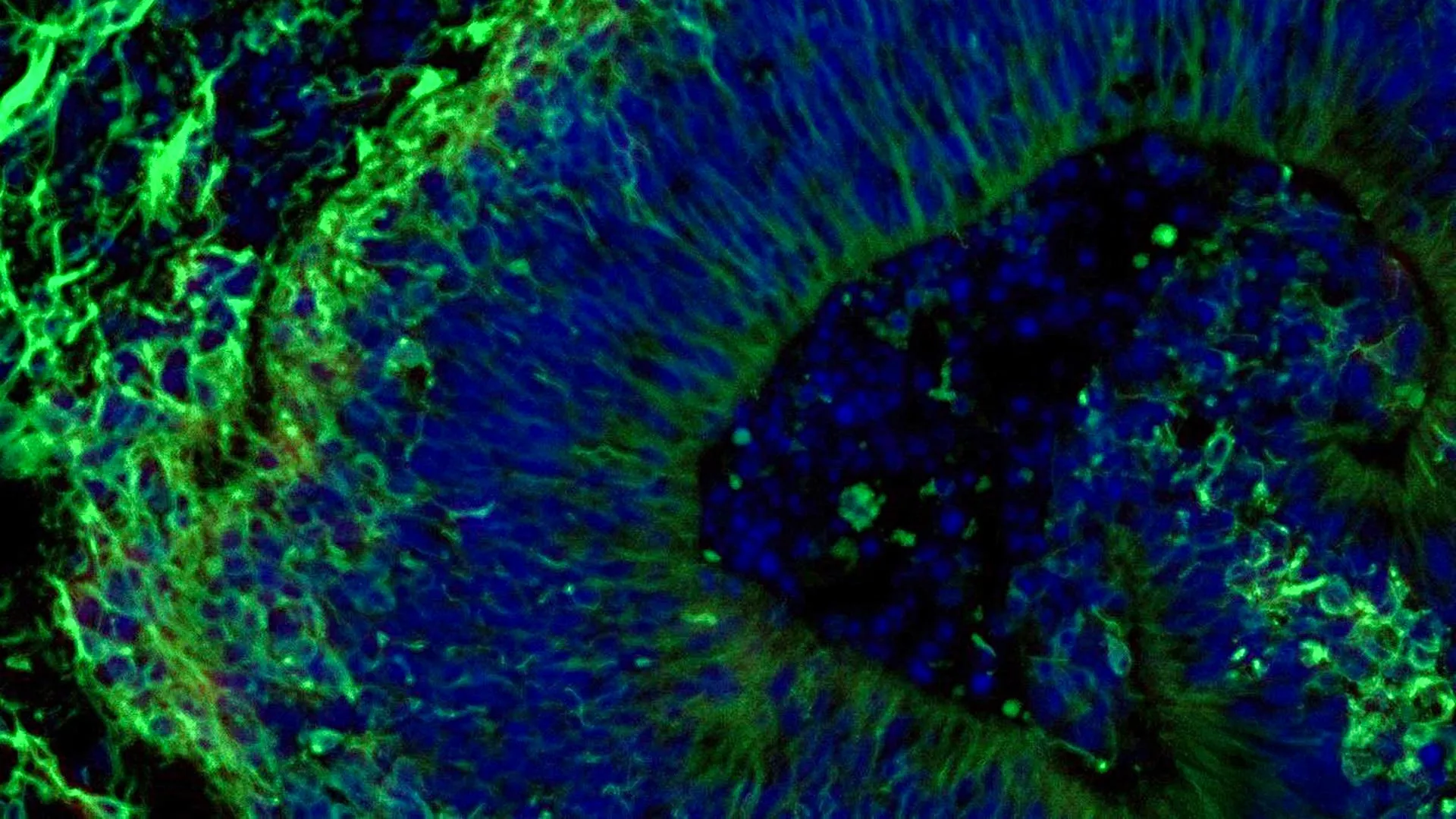
Scientists at Johns Hopkins Medicine have unveiled groundbreaking research utilizing miniature brain models, known as organoids, derived from the cells of individuals with Alzheimer’s disease. This innovative approach offers a powerful new avenue for predicting how different patients might respond to medications used to manage the complex psychiatric symptoms associated with the devastating neurodegenerative condition. The findings, published in Alzheimer’s & Dementia: The Journal of the Alzheimer’s Association, represent a significant step towards personalized medicine for Alzheimer’s disease, a condition that affects over 7 million Americans and currently has no cure.
Unlocking Personalized Treatment Strategies Through Brain Organoids
The study centers on laboratory-grown brain tissues, or organoids, meticulously developed from reprogrammed blood cells of both Alzheimer’s patients and healthy individuals. These organoids, remarkably resembling human brain tissue in their cellular composition and molecular activity, have demonstrated the capacity to mirror key biological characteristics of Alzheimer’s disease. This research builds upon a growing body of evidence suggesting that these "mini-brains" can serve as invaluable tools for understanding disease mechanisms, identifying patient subgroups, and ultimately, tailoring treatments for more effective outcomes.
"Our study suggests that large-scale, patient-derived brain organoids and the vesicles they secrete can help us stage Alzheimer’s disease, investigate the mechanisms that drive it and assess how patient subgroups may respond to different treatments," stated Dr. Vasiliki Machairaki, the study’s lead investigator and an associate professor of genetic medicine at Johns Hopkins University School of Medicine. This sentiment underscores the potential of organoid technology to move beyond a one-size-fits-all approach to managing Alzheimer’s, a disease characterized by a wide spectrum of cognitive decline and behavioral changes.
Addressing the Neuropsychiatric Burden of Alzheimer’s
Alzheimer’s disease, the most prevalent form of dementia, inflicts profound cognitive impairment, memory loss, and personality changes. Beyond these core symptoms, a significant proportion of individuals with Alzheimer’s also experience debilitating neuropsychiatric symptoms, including anxiety, depression, agitation, and psychosis. These symptoms not only diminish the quality of life for patients but also place an immense burden on caregivers and healthcare systems.
While there is no cure for Alzheimer’s, medications such as selective serotonin reuptake inhibitors (SSRIs) are frequently prescribed to alleviate these neuropsychiatric manifestations. However, the efficacy of these drugs can vary dramatically among individuals. This variability poses a significant challenge in clinical practice, often leading to trial-and-error approaches to find the most effective treatment. The Johns Hopkins research aims to address this critical unmet need by providing a predictive model for drug response.
The Genesis of the Study: A Timeline of Innovation
The research journey began with the collection of blood samples from consenting individuals diagnosed with Alzheimer’s disease at the NIH-funded Johns Hopkins Alzheimer’s Disease Research Center. This initiative, part of ongoing efforts to better understand and combat Alzheimer’s, provided the foundational material for the study.
Early 2020s (Inferred Timeline): The Johns Hopkins team embarked on the complex process of cell reprogramming. Blood cells, typically specialized and committed to their function, were meticulously reverted to an induced pluripotent stem cell (iPSC) state. This remarkable feat of cellular engineering allows these iPSCs to differentiate into virtually any cell type in the body, offering a versatile platform for disease modeling.
Mid-2020s (Inferred Timeline): Using these iPSCs, researchers then focused on generating hindbrain organoids. The hindbrain, a crucial region of the brainstem, is responsible for fundamental life-sustaining functions such as breathing, sleep-wake cycles, and heart rate regulation. The team’s decision to model this specific region was driven by its involvement in regulating mood and arousal, which are often disrupted in Alzheimer’s.
Late 2020s (Inferred Timeline): The creation of hundreds of organoids, representing a diverse cohort of Alzheimer’s patients and healthy controls, marked a significant milestone. This large-scale approach, potentially one of the most extensive brain organoid studies conducted in Alzheimer’s research to date, provided the statistical power necessary to identify subtle molecular differences and predict treatment responses.
Constructing Miniature Brains: From Blood to Organoids
The process of transforming blood cells into functional brain tissue is a testament to advances in stem cell technology and developmental biology. After obtaining blood samples, the researchers employed established protocols to reprogram somatic cells into iPSCs. These iPSCs were then coaxed into differentiating into neural progenitor cells, which were subsequently guided to self-organize into three-dimensional, pea-sized clusters that mimic the structure and cellular diversity of the human hindbrain.
Crucially, these organoids incorporated specialized brain cells, known as neurons, capable of producing serotonin. Serotonin is a key neurotransmitter that plays a vital role in regulating mood, sleep, and appetite, and is a primary target of SSRI medications. By generating organoids with this specific neuronal population, the researchers created a relevant model for investigating the effects of antidepressant drugs on Alzheimer’s-related symptoms.
Unveiling Molecular Signatures of Alzheimer’s in Organoids
A key discovery of the study was that the patient-derived organoids accurately recapitulated molecular hallmarks of Alzheimer’s disease. When compared to organoids generated from healthy individuals, those grown from the cells of Alzheimer’s patients exhibited significant differences in the expression of proteins involved in:
- Neuronal Communication: Alterations in proteins essential for the efficient transmission of signals between brain cells were observed. This disruption in synaptic function is a known contributor to cognitive decline in Alzheimer’s.
- Inflammation: The organoids displayed molecular signatures indicative of neuroinflammation, a chronic inflammatory process in the brain that exacerbates neuronal damage and disease progression.
- Disease-Specific Pathways: Proteins implicated in the pathogenesis of Alzheimer’s disease, such as those involved in amyloid plaque and tau tangle formation (though not directly modeled in this specific hindbrain organoid study, the general pathways are relevant), showed altered expression patterns.
These molecular discrepancies highlight the organoids’ fidelity as disease models, providing a tangible platform to study the complex cellular and molecular underpinnings of Alzheimer’s.
Testing the Waters: SSRI Response in Organoid Models
To assess the potential of these organoids in predicting drug response, the researchers treated a subset of the Alzheimer’s-derived organoids with escitalopram oxalate, a widely prescribed SSRI antidepressant. The results were illuminating and provided the core evidence for the study’s central hypothesis.
In some of the patient-derived organoids, the administration of escitalopram led to an increase in specific proteins associated with serotonin signaling and enhanced communication between brain cells. These are precisely the pathways that SSRIs are designed to modulate, suggesting a positive therapeutic effect. However, other organoids from different patients showed a markedly different response, exhibiting little to no molecular change after treatment.
"We used these organoids to model how some patients’ tissue may respond to a commonly prescribed SSRI," explained Dr. Machairaki. "On a large-scale level, our model may eventually be used to identify subgroups of patients, based on underlying molecular mechanisms, who are more likely to respond to certain drugs and thus help us to create precise, targeted treatments in the long run." This variability in response within the organoid models directly mirrors the clinical observation of differential drug efficacy in human patients.
Extracellular Vesicles: A New Frontier in Biomarker Discovery
Beyond analyzing the organoids themselves, the research team delved into the potential of extracellular vesicles (EVs) – tiny, membrane-bound particles released by cells – as diagnostic and prognostic tools. These vesicles act as miniature delivery systems, carrying a cargo of proteins, RNA, and other molecules that reflect the state of the parent cell.
The study found that both Alzheimer’s-derived and healthy control organoids released EVs. Upon analysis of the proteins contained within these EVs, researchers identified distinct molecular profiles. Specifically, organoids from individuals with Alzheimer’s disease showed reduced levels of proteins such as RAB3A, NSF, and ATCAY. These proteins are crucial for the normal functioning of synaptic vesicles, which are essential for neurotransmitter release and synaptic transmission. The diminished presence of these proteins in EVs from Alzheimer’s organoids suggests a potential early indicator of impaired neuronal communication.
Furthermore, the EVs released by organoids after escitalopram treatment revealed dynamic changes in protein content. In organoids that showed a molecular response to the antidepressant, the EVs contained increased levels of proteins linked to serotonin signaling and synaptic pathways. This observation suggests that EVs could potentially serve as a real-time indicator of drug efficacy, allowing clinicians to gauge a patient’s response to treatment without invasive procedures.
"This variation raises the possibility that extracellular vesicles from brain organoids could eventually help identify which patients are most likely to benefit from a particular treatment," Dr. Machairaki noted. This discovery opens up exciting possibilities for the development of non-invasive "liquid biopsies" for Alzheimer’s disease.
The Road Ahead: Enhancing Organoid Realism and Clinical Translation
While the current findings are highly promising, Dr. Machairaki and her team are committed to further refining their organoid models. Future research aims to incorporate additional cell types, such as microglia (the brain’s immune cells) and endothelial cells (which form blood vessels), into the organoids. By creating more complex and vascularized brain models, researchers can more accurately simulate the intricate environment of the living human brain, leading to even more robust and predictive experimental outcomes.
The ultimate goal is to leverage these advanced organoids and their secreted EVs to develop a comprehensive diagnostic and therapeutic platform. Such a platform could potentially:
- Diagnose Alzheimer’s disease earlier: By identifying specific EV biomarkers, clinicians might be able to detect the disease in its nascent stages, before significant cognitive decline occurs.
- Determine disease progression: Changes in EV composition could provide a quantitative measure of disease severity, allowing for more precise monitoring over time.
- Predict individual drug responses: As demonstrated in this study, organoid-derived EVs could guide treatment selection, ensuring patients receive the most effective medications for their specific disease profile.
- Identify novel therapeutic targets: Studying the molecular mechanisms revealed by organoids and EVs could uncover new avenues for drug development.
Dr. Machairaki emphasized that the current study represents an early but critical step towards achieving these ambitious goals. The collaborative efforts of researchers from Johns Hopkins, Tymora Analytical Operations, and the University of Rochester School of Medicine and Dentistry, supported by substantial funding from the National Institutes of Health and other foundations, underscore the global commitment to finding solutions for Alzheimer’s disease.
Broader Implications and Future Directions
The implications of this research extend far beyond the immediate clinical application for Alzheimer’s. The successful development and utilization of patient-derived organoids for predicting drug response represent a paradigm shift in how complex neurological disorders can be studied and treated. This approach holds promise for a wide range of conditions characterized by significant inter-patient variability, including Parkinson’s disease, epilepsy, and psychiatric disorders.
The ability to create personalized cellular models that mimic disease states offers an unprecedented opportunity to accelerate drug discovery and development. Pharmaceutical companies could potentially screen new drug candidates on organoids derived from diverse patient populations, identifying those most likely to succeed in clinical trials and reducing the high failure rates that have plagued Alzheimer’s drug development.
As the field progresses, the ethical considerations surrounding the use of human-derived cells and the interpretation of complex genomic and proteomic data will undoubtedly come to the forefront. However, the potential to alleviate the suffering caused by Alzheimer’s disease and improve the lives of millions worldwide makes this line of research exceptionally compelling and worthy of continued investment and exploration. The journey from a blood sample to a predictive model for Alzheimer’s treatment is a testament to human ingenuity and the relentless pursuit of scientific advancement.
