
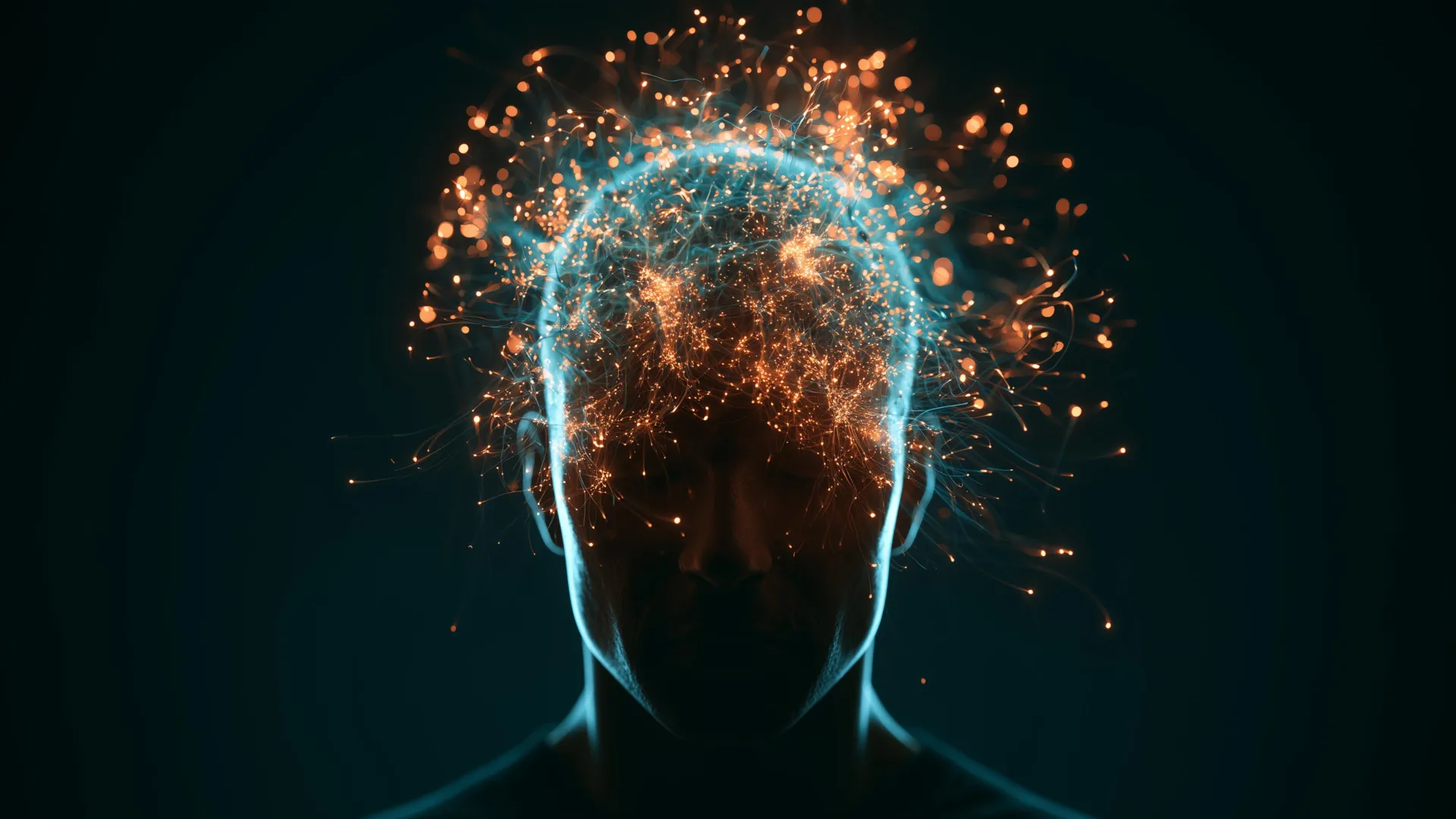
For decades, the standard medical and scientific consensus regarding general anesthesia has rested on a relatively straightforward premise: when a patient is fully unconscious under surgical-grade anesthetics, the higher-order cognitive functions of the brain are effectively suspended. While the brain stem and autonomic nervous system continue to regulate vital functions such as respiration and heart rate, the subjective experience of the world—alongside complex processing tasks like language comprehension, memory formation, and critical reasoning—is presumed to vanish entirely.
However, groundbreaking research published in the prestigious journal Nature by a team of neuroscientists and neurosurgeons at Baylor College of Medicine has shattered these traditional assumptions. The findings reveal that even when a patient is completely unresponsive and devoid of conscious awareness under general anesthesia, the brain continues to perform remarkably sophisticated cognitive feats. It can categorize complex linguistic elements, recognize subtle environmental patterns, engage in basic neural plasticity, and actively anticipate the semantic trajectory of unfolding speech.
This discovery not only forces a comprehensive reassessment of the boundary line between consciousness and unconsciousness but also opens up promising new avenues for medical technology, including the refinement of speech prosthetics and advanced brain-computer interfaces for patients suffering from severe neurological trauma.
Unprecedented Insights from Epilepsy Surgery and Neuropixels Technology
The path to this discovery began within the highly specialized environment of neurosurgical operating rooms at Baylor St. Luke’s Medical Center. Researchers led by Dr. Sameer Sheth, professor and Cullen Foundation Endowed chair of neurosurgery and a McNair Scholar at Baylor, seized a rare clinical opportunity: patients undergoing specialized surgical treatments for drug-resistant epilepsy. These surgical interventions often require direct access to deep brain structures, providing researchers with an ethical and medically safe window to record electrical activity directly from populations of human neurons that are otherwise inaccessible without invasive procedures.
Specifically, the research team focused their investigations on the hippocampus—a seahorse-shaped structure nestled deep within the brain’s temporal lobe that is universally recognized as a linchpin for human learning, memory consolidation, and spatial navigation.
To capture the activity of these neurons with unprecedented fidelity, the team deployed cutting-edge hardware known as Neuropixels probes. Developed through major neurotechnology engineering initiatives, these microscopic silicon probes feature hundreds of recording sites along a single, ultra-thin shank. This allows scientists to monitor the electrical firing of hundreds of individual neurons simultaneously with sub-millisecond precision. According to the research team, Neuropixels technology had never before been utilized to examine the human hippocampus in this precise manner during general anesthesia, offering a microscopic view of neural dynamics that previous macro-level imaging tools, such as standard electroencephalograms (EEGs) or functional magnetic resonance imaging (fMRI), could simply not achieve.
Chronology of the Experiments: From Simple Tones to Complex Narratives
The experimental design was structured in progressive phases, moving systematically from basic auditory processing to complex linguistic comprehension.
In the initial phase of the study, the anesthetized patients were exposed to a classic auditory oddball paradigm. The researchers played a continuous sequence of identical repeating tones, which were occasionally and unpredictably interrupted by a deviant sound—a tone possessing a different pitch or acoustic property. Despite the profound state of medically induced unconsciousness, single-unit recordings in the hippocampus demonstrated that neurons were not merely registering noise; they were actively distinguishing between the standard tones and the unexpected deviant sounds.
Even more striking was the observed evolution of this neural response over time. As the auditory experiment continued, the hippocampal neurons became increasingly adept at identifying and segregating the unusual tones from the background sequence. This adaptive shift exhibited classic signatures of neural plasticity—the fundamental biological mechanism by which neural circuits modify their operational strength through experience. In essence, basic learning and adaptation pathways appeared to function smoothly in the absence of any subjective awareness.
Encouraged by these results, the research team escalated the complexity of the stimulus in the subsequent phase. Rather than relying on meaningless auditory tones, the investigators played short, continuous narrative stories to the unconscious patients while maintaining continuous high-resolution recordings from the hippocampal Neuropixels probes.
Decoding Syntax, Semantics, and Predictive Coding
The analysis of the neural data captured during the storytelling phase yielded results that stunned the research team. The electrical firing patterns of the hippocampal neurons were not random or chaotic; instead, they exhibited distinct, structured configurations that reliably correlated with specific grammatical categories. The brain cells demonstrated differential responses to nouns, verbs, and adjectives, indicating that the unconscious hippocampus was processing meaningful semantic and syntactic features of language rather than just registering acoustic vibrations.
However, the most profound discovery of the experiment centered on the concept of predictive coding. The data revealed that neural activity within the hippocampus carried information that could accurately forecast upcoming words in a sentence. When a story was read aloud, the unconscious brain did not merely wait to process words after they were spoken; it actively anticipated how the narrative would continue based on context and linguistic patterns.
"The brain appears to anticipate what comes next in a story, even without conscious awareness," noted Dr. Sameer Sheth, who also serves as Director of The Gordon and Mary Cain Pediatric Neurology Research Foundation Laboratories within the Duncan Neurological Research Institute at Texas Children’s Hospital.
Predictive coding is widely considered a hallmark of conscious, attentive cognitive function. When a fully awake individual listens to speech, the brain continuously synthesizes syntax, grammar, and life experience to predict the speaker’s next words, minimizing the metabolic cost of processing entirely unexpected information. Finding robust evidence that this sophisticated predictive mechanism operates independently of consciousness bridges a major knowledge gap in cognitive neuroscience.
"This kind of predictive coding is something we associate with being awake and attentive, yet it’s happening here in an unconscious state," remarked Dr. Benjamin Hayden, professor of neurosurgery at Baylor and a co-investigator on the study.
Theoretical Implications and the Nature of Consciousness
The findings published in Nature complicate long-standing neuroscientific models regarding the exact function of consciousness. Traditionally, theories of consciousness have suggested that subjective awareness is the primary engine behind complex cognitive tasks like language interpretation, semantic categorization, and predictive inference.
The Baylor study suggests a more nuanced reality: while subjective awareness may be necessary for the experience of understanding, certain modular subsystems within the human brain retain the computational capacity to execute complex cognitive operations autonomously.
Dr. Vigi Katlowitz, a neurosurgery resident at Baylor and the study’s first author, emphasized that these insights force researchers to reconsider the architectural layout of the mind. Consciousness may not be a monolithic state where all brain regions either switch on together or shut down simultaneously. Instead, under general anesthesia, localized structures like the hippocampus may continue operating at a high level of computational sophistication, while the broader, long-range neural communication networks required to bind these signals into a unified conscious experience remain temporarily severed.
Parallels with Artificial Intelligence and Future Medical Applications
Beyond its theoretical implications for human neuroscience, the study establishes an intriguing conceptual bridge between biological neural networks and artificial intelligence (AI).
Modern large language models—the algorithmic foundation behind advanced generative AI chatbots—rely heavily on predictive coding principles. By analyzing massive datasets of human language, these AI architectures learn statistical patterns that allow them to predict and generate the most probable next word in a sequence. The observation that the human hippocampus utilizes a parallel predictive mechanism, even during complete unconsciousness, highlights a fundamental computational strategy shared by both biological evolution and machine learning systems. Exploring these structural similarities could eventually yield mutual insights for both neuroscience and computer science.
More immediately, the discovery holds significant promise for clinical translation, particularly in the development of advanced brain-computer interfaces (BCIs) and speech prosthetics. Millions of individuals worldwide suffer from locked-in syndrome, severe strokes, traumatic brain injuries, or neurodegenerative conditions that leave them cognitively intact or partially responsive but completely unable to speak.
By mapping the neural signatures associated with semantic processing and predictive language generation in deep brain structures, bioengineers may one day be able to capture these signals and translate them into synthesized speech via external neuroprosthetic devices.
"Can we use these signals to deploy and run a speech prosthetic for some of the parts of the brain that are damaged by stroke or injury? These are questions that we can now consider in relation to this part of the brain," Dr. Katlowitz stated.
Boundaries, Limitations, and the Road Ahead
While the implications of the Baylor study are sweeping, the researchers are careful to outline the technical and methodological boundaries of their work. The investigation was conducted using a specific type of general anesthetic agent administered during epilepsy surgeries, meaning the findings cannot yet be automatically generalized to all anesthetic drugs, natural slow-wave sleep, or pathological comas—states of unconsciousness that possess distinct neurophysiological profiles.
Furthermore, the study focused primarily on the hippocampus. Although the hippocampus is a central hub for memory and language integration, it operates within an extensive, distributed network of cortical and subcortical regions. Future research will need to determine whether similar sophisticated predictive and linguistic processing capabilities persist across other areas of the cerebral cortex while a patient is anesthetized.
Despite these caveats, the research marks a paradigm shift in how modern medicine views the resting or anesthetized human mind. It demonstrates that the brain behind the scenes is relentlessly active, perpetually parsing patterns, interpreting language, and anticipating the future—long before the patient opens their eyes and returns to conscious awareness.
"This work pushes us to rethink what it means to be conscious," Dr. Sheth concluded. "The brain is doing much more behind the scenes than we fully understand."
