
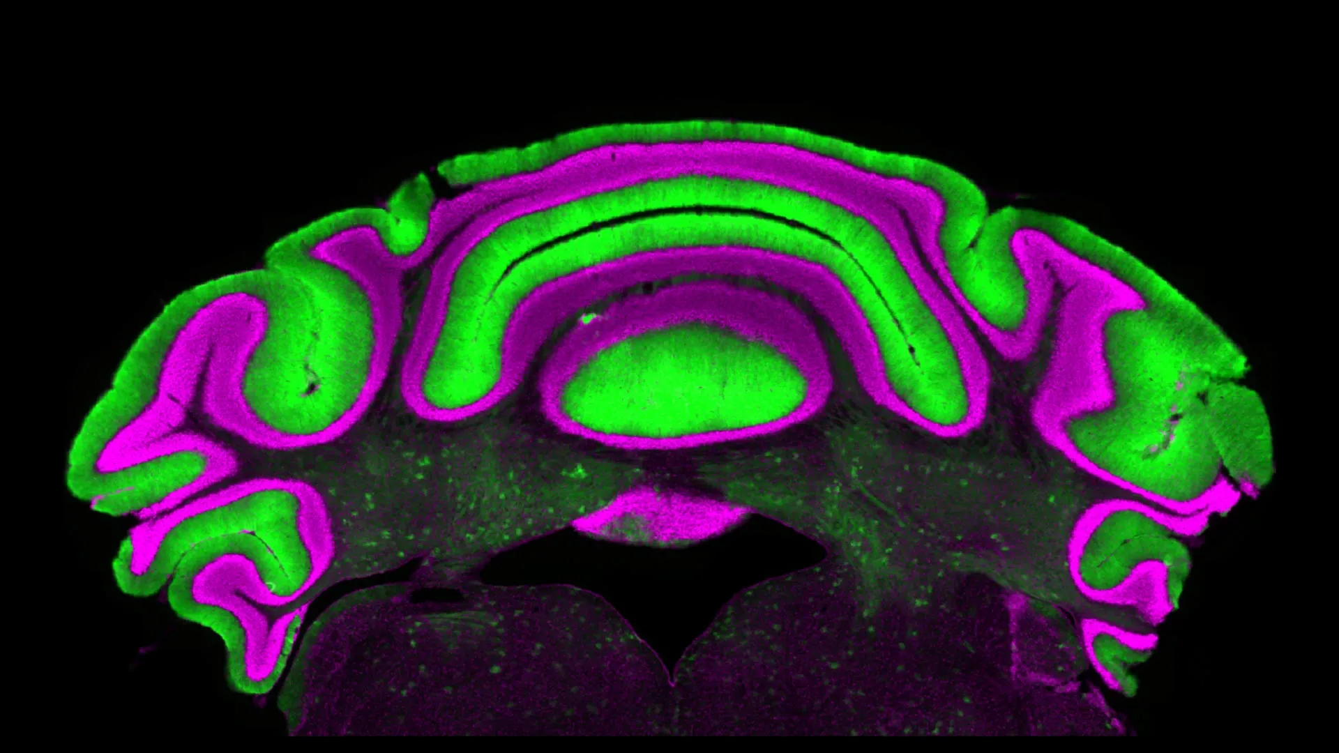
New research from a Virginia Tech neuroscientist at the Fralin Biomedical Research Institute at VTC is raising questions about a long-standing approach to studying chronic neurological conditions such as dystonia, ataxia, and tremor. For decades, the scientific community has operated under a significant assumption regarding the intricate communication within the cerebellum, a brain region critical for motor control. This assumption, that the activity of one type of nerve cell reliably mirrors the activity of another, has guided research and therapeutic development. However, the groundbreaking work by Dr. Meike van der Heijden and her team suggests this foundational understanding may be fundamentally flawed, potentially necessitating a paradigm shift in how these debilitating conditions are investigated and treated.
The cerebellum, a densely packed structure located at the back of the brain, plays an indispensable role in coordinating voluntary movements, maintaining balance, and learning motor skills. When this delicate circuitry is disrupted, individuals can experience a spectrum of debilitating symptoms. Dystonia, for instance, manifests as involuntary, sustained muscle contractions leading to abnormal postures and twisting movements. Ataxia is characterized by a lack of voluntary coordination of muscle movements, resulting in jerky, imprecise actions and gait disturbances. Tremor, a common symptom across various neurological conditions, involves rhythmic, involuntary oscillations of a body part, often the hands. These conditions, while presenting with distinct clinical features, often share a common origin in cerebellar dysfunction.
Historically, the focus of cerebellar research has largely centered on the intricate interplay between two prominent cell types: Purkinje cells and deep cerebellar nuclei (DCN) cells. Purkinje cells, the largest neurons in the brain, are known for their extensive dendritic trees, which receive a massive influx of information from other brain regions. These cells exert a powerful inhibitory influence on the DCN cells, which serve as the primary output pathway of the cerebellum, projecting to various motor control centers in the brainstem and thalamus. Given this direct inhibitory connection, the prevailing scientific consensus has been that the electrical activity of Purkinje cells serves as a reliable proxy for the activity within the DCN. Researchers have often monitored Purkinje cell activity, assuming it accurately reflects the state of the DCN, which are physically located deeper within the cerebellar cortex and are thus more challenging to access for electrophysiological recordings. This accessibility difference has further cemented the reliance on Purkinje cell data as a surrogate marker for cerebellar output.
This long-held assumption is now being directly challenged by a new study led by Dr. Meike van der Heijden, an assistant professor at the Fralin Biomedical Research Institute at VTC. Published in the esteemed Journal of Physiology, the research presents compelling evidence that the activity of Purkinje cells does not reliably predict the activity of DCN cells, even with their direct anatomical and functional connection. The study’s findings suggest that the predictive power of monitoring one cell type to understand the other is far more limited than previously believed.
Unveiling the Disconnect: A Deeper Look at Cerebellar Cell Activity
Dr. van der Heijden articulated the core finding with clarity: "We see that there’s not a clear linear relationship between activity in the Purkinje cells and in the deep nuclei cells. So there’s very limited predictive power in monitoring one to understand what’s going on in the other." This statement underscores a fundamental reevaluation of how cerebellar function is understood, particularly in the context of disease.
The research team meticulously analyzed a comprehensive database of electrophysiology recordings. These recordings were gathered from pre-clinical models exhibiting various forms of cerebellar disease, providing a crucial window into how these neural circuits behave under pathological conditions. The conventional model posited that Purkinje cells, by inhibiting DCN cells, would exhibit an inverse relationship in their activity. That is, increased Purkinje cell firing should correlate with decreased DCN cell firing, and vice versa. However, the analysis of the extensive dataset revealed a striking absence of a significant correlation between the activity patterns of these two crucial neuronal populations. This unexpected outcome directly contradicts the established paradigm that has guided cerebellar research for years.
Implications for Dystonia, Ataxia, and Tremor: A Paradigm Shift in Treatment
The implications of these findings are profound, potentially reshaping both the trajectory of scientific inquiry and the development of therapeutic interventions for a range of chronic neurological conditions. Alyssa Lyon, a doctoral candidate in Virginia Tech’s Translational Biology, Medicine, and Health Graduate Program and the paper’s first author, emphasized the clinical relevance: "Purkinje and cerebellar deep nuclei cell activity is disrupted in a disease state, and a better understanding of the relationship between these neuron types will ultimately help optimize treatments for diseases such as dystonia, ataxia, and tremor."
The long-standing reliance on Purkinje cell activity as a surrogate for DCN function stemmed, in part, from practical considerations. Purkinje cells are situated in the outermost layer of the cerebellar cortex, making them relatively more accessible for experimental manipulation and recording using techniques like electrophysiology and optical imaging. In contrast, the DCN cells are embedded deeper within the cerebellum, posing greater technical challenges for direct measurement. This disparity in accessibility inadvertently led to a situation where data from the more easily studied Purkinje cells became the de facto standard for inferring the state of the entire cerebellar output pathway.
The new research suggests that this convenience may have come at the cost of accuracy. If Purkinje cell activity does not reliably reflect DCN activity, then therapeutic strategies designed to modulate Purkinje cells with the expectation of a predictable downstream effect on DCN function may be misdirected. This could explain why some treatment approaches have yielded limited or inconsistent results.
Dr. van der Heijden elaborated on this cautionary aspect, stating, "This is a cautionary tale for understanding cerebellar activity in disease, but also for treating these challenging diseases. We need to be very careful in making assumptions, and to actually do experiments to test our hypotheses." This highlights a critical need for a more direct and nuanced approach to understanding cerebellar disorders, one that prioritizes direct measurement of DCN activity when assessing disease states and evaluating treatment efficacy.
A Historical Context: The Evolution of Cerebellar Research
The study of the cerebellum has a rich and complex history, dating back to the late 19th century. Early pioneers like Santiago Ramón y Cajal meticulously described the cellular architecture of the cerebellum, identifying its unique neuronal types, including the Purkinje cell. His groundbreaking work laid the foundation for understanding the cerebellum as a critical component of the motor system. Over the ensuing decades, advancements in neuroscience techniques allowed for increasingly sophisticated investigations into cerebellar function.
The discovery of the inhibitory nature of Purkinje cells by Rodolfo Llinás in the mid-20th century was a pivotal moment, solidifying the inhibitory Purkinje cell-excitatory DCN neuron circuit as a central tenet of cerebellar physiology. This model, while influential, has largely been explored through the lens of Purkinje cell activity due to experimental accessibility.
The emergence of sophisticated genetic tools, advanced microscopy, and refined electrophysiological recording methods in recent decades has opened new avenues for studying neural circuits with unprecedented detail. It is within this context of advanced technological capabilities that Dr. van der Heijden’s team has been able to revisit and rigorously test long-standing assumptions. The study’s methodology, which involved analyzing large datasets of pre-clinical electrophysiological recordings, represents a powerful form of meta-analysis, leveraging existing data to uncover novel insights.
Future Directions and Therapeutic Implications
The ramifications of this research extend beyond academic curiosity, offering tangible hope for improved clinical outcomes. By revealing the dissociation between Purkinje cell and DCN activity, the study directs future research efforts towards more direct investigations of DCN function in disease. This could involve the development of more refined techniques for recording from and manipulating DCNs in both animal models and, eventually, in human patients.
For conditions like dystonia, ataxia, and tremor, where cerebellar dysfunction is a known contributor, a more accurate understanding of the underlying cellular mechanisms is paramount. If treatments aimed at modulating Purkinje cells are not effectively translating to the DCN, the target of therapeutic interventions may need to be re-evaluated. This could lead to the exploration of novel drug targets or neuromodulation strategies that directly influence DCN activity.
Furthermore, the study underscores the importance of robust experimental validation. The "cautionary tale" that Dr. van der Heijden highlights is a call to action for the broader neuroscience community to rigorously test hypotheses, even those that have become deeply ingrained in the field. The scientific method thrives on skepticism and the willingness to revise established paradigms when confronted with compelling evidence.
The research team’s commitment to open science and data sharing, as evidenced by the analysis of existing databases, also exemplifies a modern approach to scientific discovery. By building upon the work of previous researchers, they have been able to advance the field in a significant way.
Broader Impact on Neurological Research
The implications of this Virginia Tech study resonate across the broader landscape of neurological research. The cerebellum is implicated in a wider range of cognitive and emotional functions than previously appreciated, including learning, language, and social behavior. The intricate circuits within the cerebellum are also thought to play a role in conditions beyond movement disorders, such as autism spectrum disorder and schizophrenia. Therefore, a more accurate understanding of cerebellar cell communication could have far-reaching consequences for research into a diverse array of neurological and psychiatric conditions.
The challenge now lies in translating these fundamental discoveries into clinical practice. This will require collaborative efforts between basic scientists, clinicians, and pharmaceutical companies. The development of new diagnostic tools that can accurately assess DCN activity in patients, alongside the creation of targeted therapeutic interventions, will be crucial steps forward.
In conclusion, the research conducted at the Fralin Biomedical Research Institute at VTC represents a significant leap forward in our understanding of cerebellar function. By challenging a long-held assumption, Dr. van der Heijden and her team have opened new avenues for research and treatment development, offering renewed hope for individuals affected by chronic neurological conditions like dystonia, ataxia, and tremor. The findings serve as a powerful reminder that scientific progress often hinges on the courage to question established beliefs and the dedication to rigorous empirical investigation.
