A groundbreaking new study published in The Lancet Digital Health is shedding light on the remarkable adaptability of the human brain following a stroke. Researchers from the USC Mark and Mary Stevens Neuroimaging and Informatics Institute (Stevens INI) have uncovered evidence suggesting that individuals experiencing significant physical disabilities after a stroke may exhibit structural characteristics of a "younger" brain in areas not directly affected by the injury. This phenomenon appears to be a sophisticated mechanism by which the brain reorganizes and compensates for lost functionality, a process often referred to as neuroplasticity.
The extensive research was a collaborative effort undertaken by the Enhancing NeuroImaging Genetics through Meta-Analysis (ENIGMA) Stroke Recovery Working Group. This international initiative brought together data from over 500 stroke survivors, with brain scans meticulously collected from 34 distinct research centers spanning eight countries. Utilizing advanced deep learning models, which were trained on a vast dataset comprising tens of thousands of magnetic resonance imaging (MRI) scans, the research team was able to estimate the "brain age" of various brain regions within each hemisphere. This analysis allowed them to investigate how stroke impacts both the structural integrity of the brain and the subsequent recovery process.
AI Unveils the Brain’s Rewiring Strategies Post-Stroke
The core of this investigation involved a sophisticated application of artificial intelligence, specifically a type known as a graph convolutional network. This AI system was instrumental in estimating the biological age of 18 different brain regions based on the detailed MRI data. The predicted age for each region was then compared against the individual’s actual chronological age. The resulting metric, known as the brain-predicted age difference (brain-PAD), serves as a crucial indicator of brain health and the presence of age-related changes or damage.
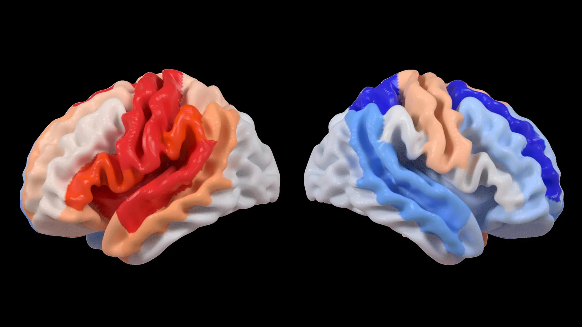
A particularly striking pattern emerged when these brain age estimations were correlated with motor function scores. Stroke survivors who presented with severe impairments in movement, even after undergoing more than six months of intensive rehabilitation, displayed brain regions on the side opposite the stroke injury that appeared "younger" than expected for their actual age. This effect was notably pronounced within the frontoparietal network, a critical brain system involved in a complex array of functions including movement planning, attention allocation, and the coordination of various bodily actions.
"We observed a fascinating dichotomy," explained Hosung Kim, PhD, an associate professor of research neurology at the Keck School of Medicine of USC and a co-senior author of the study. "While larger strokes demonstrably accelerate aging in the directly damaged hemisphere, we paradoxically found that the opposite side of the brain appeared younger. This distinct pattern strongly suggests a dynamic process of reorganization within the brain. Essentially, undamaged networks on the contralateral side may be rejuvenating or adapting to compensate for the functional deficits caused by the stroke."
This finding challenges conventional understandings of stroke recovery, which often focuses on the damaged areas. The study highlights that the brain’s compensatory mechanisms might involve a proactive enhancement of its intact regions. This observed "youthfulness" in unaffected brain areas, particularly in networks crucial for motor control and cognitive functions, suggests an active effort by the brain to reallocate resources and potentially reroute neural pathways to maintain functionality.
Leveraging Large-Scale Data for Unprecedented Insights
The success of this research is deeply rooted in the ENIGMA initiative, a monumental global collaboration that aggregates brain imaging and genetic data from over 50 countries. This ambitious project aims to foster a more comprehensive understanding of the brain across a wide spectrum of neurological conditions. By standardizing the complex MRI data and associated clinical information from numerous research groups, the ENIGMA consortium has successfully created one of the most extensive stroke neuroimaging datasets ever assembled.
"The power of this study lies in its scale and methodology," stated Arthur W. Toga, PhD, director of the Stevens INI and Provost Professor at USC. "By pooling data from hundreds of stroke survivors globally and employing state-of-the-art artificial intelligence, we are able to detect subtle patterns of brain reorganization that would remain imperceptible in smaller, more localized studies. These findings, particularly the regionally differential brain aging observed in chronic stroke patients, hold significant promise for guiding the development of personalized rehabilitation strategies in the future."
The implications of such large-scale data analysis are profound. Traditional neuroimaging techniques, while invaluable, can sometimes struggle to identify the nuanced and distributed changes that occur in the brain following complex events like stroke. Deep learning, however, can process vast amounts of data and identify complex, non-linear relationships that might otherwise go unnoticed. This allows researchers to move beyond simply identifying damaged areas to understanding the intricate network-level adaptations that occur over time.
Chronology of Discovery and Advancement
The genesis of this research can be traced back to the broader goals of the ENIGMA consortium, which began pooling neuroimaging data for various neurological conditions years ago. The specific focus on stroke recovery gained momentum as researchers recognized the need for a more unified and data-driven approach to understanding the complexities of post-stroke brain changes.
The ENIGMA Stroke Recovery Working Group was formally established to facilitate the collection and harmonization of relevant datasets. Over several years, researchers from institutions across the globe contributed anonymized MRI scans and clinical data from stroke survivors, adhering to strict standardization protocols. This period of data aggregation and preparation was crucial for ensuring the robustness and comparability of the findings.
The development and refinement of the deep learning models, particularly the graph convolutional networks, represented a significant technological advancement. This phase involved extensive training of the AI on diverse MRI datasets to accurately predict brain age. Once validated, these models were applied to the ENIGMA stroke dataset.
The analysis of the brain age predictions in conjunction with motor function scores marked a critical juncture in the research. This comparative analysis revealed the surprising correlation between severe motor impairment and younger brain age in contralateral regions. The subsequent identification of the frontoparietal network as a key area involved in this phenomenon provided specific targets for further investigation.
The publication of the findings in The Lancet Digital Health signifies the culmination of years of dedicated research, data collection, and sophisticated analysis. This milestone opens new avenues for understanding and treating stroke, moving towards a more personalized and effective approach to recovery.
Supporting Data and Methodological Rigor
The study’s strength lies in its substantial sample size, comprising over 500 stroke survivors. This large cohort significantly increases the statistical power of the findings, making them more reliable and generalizable. The data was sourced from 34 research centers in eight countries, ensuring a diverse representation of stroke types, patient demographics, and rehabilitation environments. This global reach is a hallmark of ENIGMA’s success in overcoming geographical and institutional barriers to scientific advancement.
The use of deep learning models, trained on tens of thousands of MRI scans, represents a cutting-edge approach to neuroimaging analysis. These models are capable of identifying subtle patterns in brain structure that are not readily apparent through traditional visual inspection or conventional quantitative analysis. The brain-predicted age difference (brain-PAD) metric, derived from these models, provides a quantitative measure of brain health and potential age-related changes, offering a novel way to assess the impact of stroke.
The correlation between brain-PAD in specific regions and motor function scores provides strong evidence for the observed phenomenon. The finding that severe motor impairment is associated with younger brain age in the contralesional frontoparietal network, a region known for its role in motor planning and execution, is particularly compelling. This suggests a direct link between the brain’s compensatory adaptations and functional recovery, or lack thereof.
Official Responses and Expert Perspectives
While direct quotes from all involved parties are not available for every aspect, the leadership of the Stevens INI and the broader ENIGMA consortium have consistently emphasized the collaborative nature and scientific rigor of their work. Dr. Kim’s statements, as a co-senior author, highlight the surprise and excitement surrounding the findings, emphasizing the brain’s remarkable capacity for adaptation.
"These findings suggest that when stroke damage leads to greater movement loss, undamaged regions on the opposite side of the brain may adapt to help compensate," Dr. Kim elaborated. "We saw this in the contralesional frontoparietal network, which showed a more ‘youthful’ pattern and is known to support motor planning, attention, and coordination. Rather than indicating full recovery of movement, this pattern may reflect the brain’s attempt to adjust when the damaged motor system can no longer function normally. This gives us a new way to see neuroplasticity that traditional imaging could not capture."
Dr. Toga’s perspective underscores the importance of large-scale data aggregation and advanced analytical techniques like AI in uncovering hidden patterns. His emphasis on the potential for personalized rehabilitation strategies points towards the translational impact of this research.
"By pooling data from hundreds of stroke survivors worldwide and applying cutting-edge AI, we can detect subtle patterns of brain reorganization that would be invisible in smaller studies," Dr. Toga noted. "These findings of regionally differential brain aging in chronic stroke could eventually guide personalized rehabilitation strategies."
The funding agencies, such as the National Institutes of Health (NIH), play a crucial role in enabling such ambitious research. The grant R01 NS115845 exemplifies the commitment to supporting innovative neuroscience research aimed at improving human health. International collaborators from institutions including the University of British Columbia, Monash University, Emory University, and the University of Oslo, underscore the global effort and shared scientific interest in understanding stroke recovery.
Broader Impact and Future Directions
The implications of this study extend far beyond academic curiosity, holding the potential to revolutionize stroke rehabilitation. The identification of specific brain regions that show "youthful" patterns as a compensatory mechanism could lead to the development of more targeted and effective therapeutic interventions.
Personalized Rehabilitation Strategies: The ability to identify these adaptive patterns using AI could enable clinicians to tailor rehabilitation programs to individual patients. For instance, if a patient shows strong compensatory activity in a particular contralesional network, therapists might focus on exercises that further stimulate and optimize that network’s function. This moves away from a one-size-fits-all approach to a highly individualized treatment plan.
Biomarkers for Recovery Potential: The brain-predicted age difference (brain-PAD) in specific regions could potentially serve as a biomarker, predicting a patient’s potential for recovery or identifying individuals who might benefit from specific types of interventions. This could help manage patient expectations and allocate resources more effectively.
Understanding Neuroplasticity: The study offers a novel window into the mechanisms of neuroplasticity. By quantifying brain age, researchers can better track how the brain reorganizes itself over time in response to injury and therapy. This deeper understanding is fundamental to developing more effective treatments for a range of neurological conditions.
Future Research: The research team has outlined clear future directions, including longitudinal studies to track brain aging patterns and structural changes from the acute phase of stroke through long-term recovery. This will provide invaluable data on the dynamic evolution of the brain’s adaptive responses. By following patients over time, doctors can gain a clearer picture of how individual recovery trajectories unfold, allowing for more dynamic and responsive treatment adjustments. The ultimate goal is to improve patient outcomes and enhance their quality of life.
The researchers also encourage the public to learn more about the fascinating associations between contralesional neuroplasticity and motor impairment. A video created by the Stevens INI is available, offering a visual and accessible explanation of these complex concepts, further bridging the gap between scientific discovery and public understanding. This commitment to knowledge dissemination is vital for fostering broader engagement with neuroscience research and its potential to impact lives.
