
The assembly of the human brain before birth stands as one of the most complex biological processes in nature, relying on a delicate orchestration of cellular choices, chemical signals, and physical interactions. At the heart of this intricate developmental architecture are radial glia, a specialized category of neural stem cells. These remarkable cells are responsible for generating the vast majority of neurons and support cells that constitute the cerebral cortex—the outermost layer of the brain tasked with higher-order functions such as abstract thought, episodic memory, and complex language processing.
For decades, developmental neurobiologists have understood that radial glia play a critical role in the disproportionate evolutionary expansion of the human cerebral cortex compared to other mammalian species. However, the precise molecular and cellular mechanisms that dictate how these master stem cells decide to divide, differentiate, or transform have remained largely elusive. Furthermore, while most radial glia naturally disappear before birth, scientists have observed that similar cellular programs can reactivate years later in aggressive brain cancers, operating through pathways that researchers are only beginning to decode.
Now, a pair of landmark studies published concurrently in the prestigious journals Cell and Science has provided unprecedented insight into the inner workings of radial glia. Led by researchers at the University of California, Los Angeles (UCLA), these investigations reveal that radial glia do not make their developmental decisions in isolation. Instead, their cellular fates are actively steered by two distinct and unexpected external inputs: metabolic processing within the cellular microenvironment and direct physical signals originating from deep-seated structures within the developing brain.
Decoding the Cellular Architects of the Human Mind
To appreciate the significance of these findings, researchers must examine the unique evolutionary trajectory of the human brain. The cerebral cortex has expanded dramatically over millions of years of human evolution, a phenomenon largely driven by the prolonged proliferative capacity of radial glia during gestation. These cells act as biological factories, producing wave after wave of neurons that migrate outward to construct the layered architecture of the cortex.
Despite their central importance, studying human radial glia has historically presented formidable technical hurdles. Because human brain development occurs entirely in utero and cannot be observed directly in living subjects, scientists have traditionally relied on animal models, primarily rodents. However, rodent brains lack the massive cortical expansion, structural complexity, and specific cell-type diversity characteristic of humans. Consequently, many pathways governing human-specific cortical development could not be reliably studied in traditional laboratory animals.
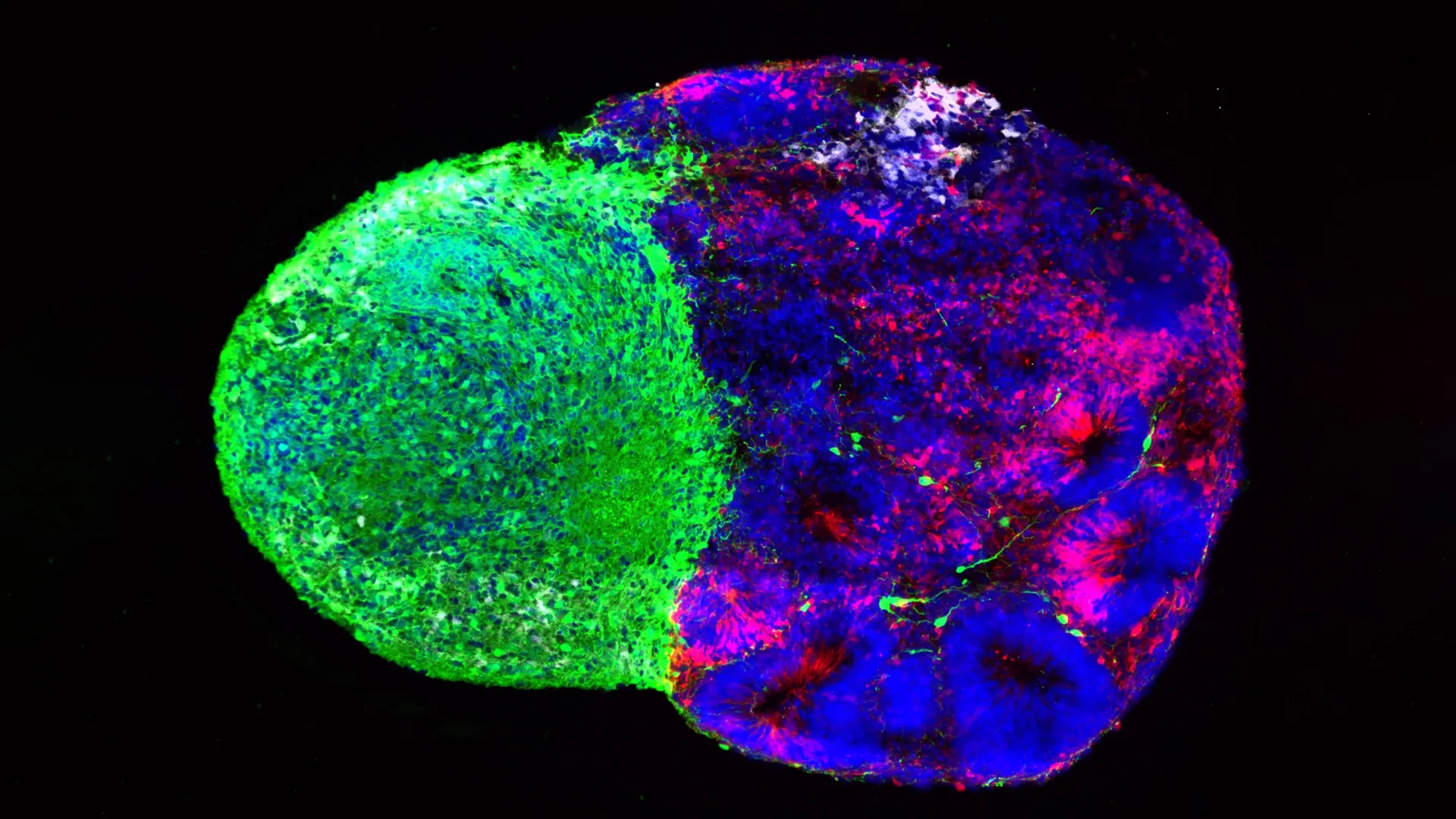
The landscape of neurodevelopmental research shifted dramatically over the past decade with the advent of advanced stem cell technologies, including human brain organoids and multi-cellular "assembloids." These laboratory-grown tissue models replicate key structural and functional features of the developing human brain, enabling scientists to observe human neural stem cells directly in real time. Capitalizing on these technological breakthroughs, a collaborative team of UCLA researchers set out to investigate the upstream drivers of radial glia behavior, resulting in the twin breakthroughs recently published in Cell and Science.
Metabolism as an Active Driver of Cellular Fate
In the first of the two studies, published in Cell, researchers sought to understand how cellular metabolism influences the developmental trajectory of neural stem cells. Spearheaded by co-first authors Jessenya Mil and Jose Soto, the project was a collaborative effort between the laboratory of Dr. Aparna Bhaduri—an assistant professor of biological chemistry at the David Geffen School of Medicine at UCLA—and the laboratory of Dr. Heather Christofk.
Traditionally, cellular metabolism has been viewed by biologists as a passive background support system—a set of housekeeping chemical reactions required merely to keep cells alive and fueled with energy. However, the UCLA research team hypothesized that metabolic pathways might play a far more active, instructive role in guiding cellular differentiation during organogenesis.
To test this hypothesis, the researchers constructed a high-resolution metabolic atlas of the developing human cortex. By analyzing donated human embryonic and fetal tissue alongside sophisticated brain organoids derived from human stem cells, the team mapped metabolic activity across various stages of cortical formation.
The resulting data pointed to an unexpected conclusion. The analysis revealed that radial glia rely heavily on a specific metabolic pathway known as the pentose phosphate pathway. This biochemical route utilizes glucose to synthesize nucleic acids and other essential building blocks required by rapidly dividing cells. When the research team experimentally manipulated this pathway—either by restricting the availability of glucose or by introducing chemical inhibitors that blocked key enzymes within the pentose phosphate pathway—the behavior of the stem cells shifted dramatically.
Rather than continuing down their standard developmental pathway, the radial glia altered their output, generating higher proportions of inhibitory neurons and other specialized cell types that typically emerge later in fetal development. This demonstrated that metabolic flux is not merely a passive consequence of cell growth, but an active regulatory mechanism capable of instructing stem cells on which cellular identities to adopt.
Implications for Maternal Health and Metabolic Disorders
The identification of metabolism as a primary driver of neural stem cell differentiation opens significant new avenues for clinical research. Scientists note that these findings could help clarify how maternal health factors—such as gestational diabetes, nutritional deficiencies, obesity, and systemic inflammation—can impact fetal brain development and increase the risk of neurodevelopmental abnormalities in offspring.
Furthermore, the newly established metabolic atlas provides the scientific community with an exhaustive reference tool. Researchers studying pediatric neurological conditions, metabolic disorders, and brain tumors can utilize this comprehensive dataset to investigate how metabolic dysregulation intersects with abnormal cellular proliferation. By understanding how cancer cells hijack these same metabolic programs to fuel unchecked growth, oncologists may eventually identify novel therapeutic vulnerabilities in malignant brain tumors.
Physical Projections from the Thalamus Shape Cortical Development
While the Cell study explored the internal metabolic environment of neural stem cells, the second investigation, published in Science and led by first author Claudia Nguyen, examined external signals arriving from a completely different anatomical region of the brain.
The researchers trained their focus on the thalamus, a deeply situated relay center located near the center of the brain that processes sensory and motor signals before routing them to appropriate cortical destinations. Decades of anatomical research have established that thalamic neurons extend extraordinarily long, wire-like projections toward the cerebral cortex. These fibers eventually form intricate synaptic networks with specific cortical neurons, facilitating communication across the central nervous system.
However, detailed anatomical observations of human fetal tissue have long presented a temporal puzzle: these thalamic projections arrive at the cortex weeks or even months before the cortical neurons are fully formed and ready to receive them. For years, neuroscientists wondered why these nerve fibers migrated to the cortex so early in gestation, long before functional synaptic connections could possibly be established.
Using human stem cell-derived brain assembloids—complex laboratory models that fuse separately grown regional organoids to observe how different brain regions interact—the UCLA team uncovered a surprising answer. The research revealed that the arriving thalamic projections physically touch the radial glia cells directly while the cortex is still under construction.
Direct Cellular Contact Triggers Excitatory Neuron Production
When these physical contacts occur, they induce immediate behavioral changes in the stem cells. The direct touch stimulates radial glia to produce a higher volume of excitatory neurons, which serve as the primary signal-carrying neurons responsible for cognitive processing and information transmission in the cortex. Notably, this stimulatory effect was found to be especially pronounced for upper-layer cortical neurons—the specific neuronal populations that have undergone the most dramatic evolutionary expansion in the human brain compared to other primates and rodents.
"We already knew that these projections influence how the cortex develops," Dr. Bhaduri explained, reflecting on the significance of the observation. "What we specifically found is that this influence comes through an actual physical connection between the projections and the radial glia—a point of contact that just hasn’t been identified before, and one that very likely does not exist in rodents."
This discovery highlights a fundamental structural difference in human neurodevelopment. Because such intricate physical interactions between thalamic fibers and radial glia appear unique to primates and humans, animal models have historically failed to capture this critical developmental checkpoint. The use of human assembloids thus bridges a major translational gap in developmental neuroscience.
Linking Cellular Contact to Autism-Associated Genes
To further investigate the molecular machinery mediating this physical interaction, the UCLA researchers focused on NRXN1, a gene widely recognized for its role in helping neurons establish structural connections with one another. Genetic mutations and deletions involving the NRXN1 gene have long been strongly associated with an increased susceptibility to autism spectrum disorder and other neurodevelopmental conditions.
To determine whether NRXN1 plays a role in early stem cell signaling as well as mature synaptic formation, the team engineered human assembloids using patient-derived stem cells carrying a documented NRXN1 mutation. Upon testing these mutated models, the researchers observed that the altered thalamic signals interacted aberrantly with the radial glia.
This cellular miscommunication disrupted the tightly regulated balance between maintaining the stem cell pool and generating differentiated neurons. By demonstrating how a genetic mutation linked to autism can alter physical and chemical signaling events during the earliest phases of cortical assembly, the research provides a tangible cellular framework for investigating the origins of neurodevelopmental disorders long before birth.
A Unified View of Brain Assembly
Taken together, these two independent studies offer a cohesive paradigm shift in how neuroscientists view the construction of the human brain. Whether through the quiet chemical currents of cellular metabolism or the direct mechanical touch of distant neural projections, radial glia do not operate in a vacuum. Their developmental decisions are continuously modulated by a dense, dynamic network of environmental signals.
The publication of these findings also underscores the rapid maturation of stem cell technology over the past decade. Where researchers once faced insurmountable ethical and technical barriers in studying living human neural tissue, modern organoid and assembloid platforms now permit safe, reproducible, and highly detailed exploration of human neurogenesis in vitro.
As the scientific community digests these insights, researchers emphasize that viewing metabolism and physical cell-to-cell contact as active developmental drivers—rather than passive background conditions—will be essential for future breakthroughs. By illuminating the fundamental rules that govern how radial glia build the human mind, these studies lay vital groundwork for diagnosing neurodevelopmental disorders, understanding disease vulnerability, and ultimately decoding the cellular origins of brain cancer.
Support for this research was provided by a broad coalition of major scientific organizations and philanthropic foundations, including the National Institutes of Health, the National Science Foundation, the Brain & Behavior Research Foundation, the Alfred P. Sloan Foundation, the Rose Hills Foundation, the Esther A. & Joseph Klingenstein Fund, the Simons Foundation, the Chan Zuckerberg Initiative, the NIH BRAIN Initiative Cell Atlas Network, the International Foundation for Ethical Research, the UCLA Broad Stem Cell Research Center’s Stem Cell Research Training Program, and the UCLA Health Jonsson Comprehensive Cancer Center and UCLA Broad Stem Cell Research Center Ablon Scholars Program.
