
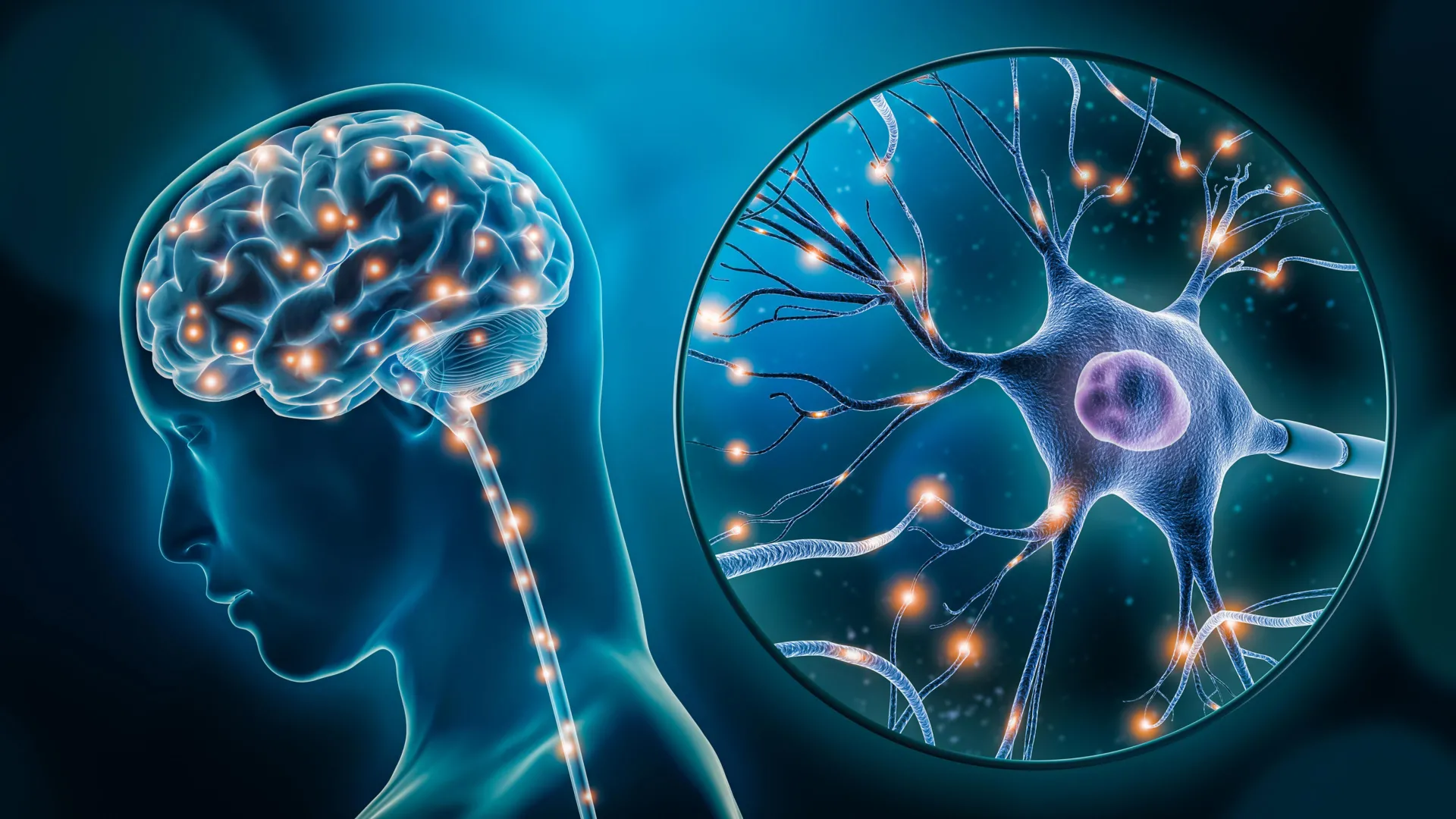
Researchers at McGill University and the Douglas Institute have unveiled a groundbreaking discovery that illuminates the intricate biological underpinnings of depression, identifying two distinct types of brain cells that function differently in individuals experiencing this pervasive mental health condition. This pivotal research, published in the esteemed journal Nature Genetics, not only offers crucial insights into the cellular mechanisms driving depression but also paves the way for the development of highly targeted therapeutic interventions. With over 264 million people worldwide affected by depression, and its status as a leading cause of global disability, these findings represent a significant leap forward in understanding and combating this complex disorder.
A Novel Approach to Unraveling Depression’s Cellular Roots
The study, led by Dr. Gustavo Turecki, a distinguished professor at McGill, a clinician-scientist at the Douglas Institute, and holder of the Canada Research Chair in Major Depressive Disorder and Suicide, marks a significant advancement in psychiatric research. "This is the first time we’ve been able to identify what specific brain cell types are affected in depression by mapping gene activity together with mechanisms that regulate the DNA code," Dr. Turecki stated. "It gives us a much clearer picture of where disruptions are happening, and which cells are involved." This innovative approach moves beyond broad observations of brain activity to pinpoint specific cellular actors and their altered genetic regulation.
The breakthrough was made possible by access to a unique and invaluable resource: post-mortem brain tissue samples from the Douglas-Bell Canada Brain Bank. This specialized repository is one of the few globally that meticulously collects and preserves donated brain tissue from individuals who had psychiatric conditions, including depression. Such samples are critical for in-depth biological investigations into mental health disorders, allowing researchers to examine the brain at a cellular and molecular level long after the individual’s passing.
Utilizing cutting-edge single-cell genomic techniques, the research team meticulously analyzed RNA and DNA from thousands of individual brain cells. This advanced methodology enabled them to not only differentiate between cell types but also to ascertain which specific cells exhibited altered behavior in individuals diagnosed with depression. The study encompassed a robust sample size, including brain tissue from 59 individuals who had a diagnosed history of depression and 41 individuals without the condition, providing a solid foundation for comparative analysis.
Identifying Key Cellular Players in Depression
The comprehensive analysis revealed significant alterations in gene activity within two crucial types of brain cells: a specific population of excitatory neurons and a subtype of microglia.
Excitatory Neurons and Mood Regulation: The first group identified involves excitatory neurons, a fundamental class of nerve cells responsible for transmitting signals throughout the brain. These neurons play a critical role in a myriad of cognitive and emotional processes, including the regulation of mood, the capacity to respond to stress, and the facilitation of learning and memory. In individuals with depression, the study observed differential gene expression within these neurons, suggesting that their ability to effectively regulate mood and manage stress responses may be compromised. This disruption could manifest as an inability to experience pleasure, persistent feelings of sadness, or an overactive stress response, all hallmarks of depressive disorders.
Microglia and Neuroinflammation: The second cell type exhibiting altered activity was a particular subtype of microglia. Microglia are the primary immune cells of the central nervous system, acting as the brain’s resident macrophages. Their crucial functions include clearing cellular debris, responding to injury or infection, and critically, modulating neuroinflammation. Chronic or dysregulated neuroinflammation has increasingly been implicated in the pathophysiology of various neurological and psychiatric conditions, including depression. The observed changes in gene activity within these microglial subtypes suggest a potential role for altered immune responses and inflammatory processes within the brain of individuals with depression. This could lead to a state of chronic, low-grade inflammation that negatively impacts neuronal function and contributes to depressive symptoms.
The simultaneous observation of altered gene activity in both these distinct cell types—neurons responsible for signal transmission and mood regulation, and immune cells involved in brain maintenance and inflammation—underscores the complex, multi-faceted biological nature of depression. These findings suggest that depression is not merely an imbalance of neurotransmitters, as once primarily theorized, but rather a disruption in intricate cellular communication and regulatory networks.
Implications for Understanding and Treating Depression
The study’s findings carry profound implications for how depression is understood and treated. By providing concrete evidence of specific cellular and genetic dysfunctions, the research reinforces the scientific consensus that depression is a legitimate brain disorder with a clear biological basis. This empirical data challenges outdated perspectives that may have relegated depression to the realm of purely psychological or emotional distress, often leading to stigma and inadequate treatment.
"This research reinforces what neuroscience has been telling us for years," Dr. Turecki emphasized. "Depression isn’t just emotional, it reflects real, measurable changes in the brain." This statement serves as a powerful affirmation for individuals living with depression and their loved ones, validating their experiences as rooted in biological realities rather than personal failing.
The identification of specific cell types and their altered genetic machinery opens up exciting new avenues for therapeutic development. Current antidepressant medications, while effective for many, often have broad mechanisms of action and can take weeks to show benefits, with significant side effects for some individuals. The ability to target specific cellular pathways within excitatory neurons or microglia could lead to the creation of more precise and potentially more effective treatments. For instance, therapies could be designed to:
- Restore normal excitatory neuron function: This might involve interventions aimed at modulating neurotransmitter signaling, improving synaptic plasticity, or enhancing the resilience of these neurons to stress.
- Regulate microglial activity: Treatments could focus on dampening excessive neuroinflammation or promoting the beneficial functions of microglia, thereby creating a more supportive environment for neuronal health.
This targeted approach holds the promise of not only improving treatment efficacy but also reducing the incidence of adverse drug reactions, thereby enhancing the quality of life for individuals undergoing treatment.
The Journey from Discovery to Clinical Application: A Timeline
The research process leading to this significant publication involved several key stages, representing a typical trajectory for scientific breakthroughs:
Initial Hypothesis and Funding Acquisition (Years Prior): Building upon decades of neuroscience research that suggested a biological component to depression, researchers like Dr. Turecki would have formulated hypotheses regarding specific cellular mechanisms. This stage would involve extensive grant writing and securing funding from governmental agencies and private foundations. The Canadian Institutes of Health Research, Brain Canada Foundation, Fonds de recherche du Québec – Santé, and the Healthy Brains, Healthy Lives initiative at McGill University provided crucial financial support for this study, underscoring the national and institutional commitment to mental health research.
Sample Collection and Preparation (Ongoing): The Douglas-Bell Canada Brain Bank has been accumulating its invaluable collection over many years, a testament to the generosity of donors and the foresight of its organizers. The preparation of these sensitive biological samples for genomic analysis is a meticulous and time-consuming process, requiring specialized expertise and sterile conditions.
Advanced Genomic Analysis (Several Years): The application of single-cell genomic techniques, such as single-nucleus chromatin accessibility profiling (ATAC-seq), is technologically demanding and computationally intensive. This phase involves extracting high-quality DNA and RNA from individual cells, performing sequencing, and then processing vast amounts of data to identify differences in gene expression and epigenetic regulation. The researchers likely spent several years refining their methods and analyzing the data from hundreds of brain samples.
Data Interpretation and Validation (Months to a Year): Once the raw data is generated, the critical phase of interpretation begins. This involves statistical analysis, biological pathway enrichment, and cross-referencing with existing literature to draw meaningful conclusions. Validation experiments, potentially using animal models or cell cultures, might also be conducted to confirm key findings, although the primary focus of this study was on human post-mortem tissue.
Manuscript Preparation and Peer Review (Several Months): The process of writing a scientific paper for a high-impact journal like Nature Genetics is rigorous. It involves clearly articulating the research question, methods, results, and implications, followed by a lengthy and stringent peer-review process where other experts in the field scrutinize the study for its scientific validity and originality. The publication of "Single-nucleus chromatin accessibility profiling identifies cell types and functional variants contributing to major depression" by Anjali Chawla and Gustavo Turecki et al. signifies the culmination of this extensive scientific endeavor.
Future Research and Clinical Translation (Ongoing): The findings presented are not an endpoint but a new beginning. The researchers’ immediate next steps involve delving deeper into how these observed cellular differences impact overall brain network function and exploring the therapeutic potential of targeting these specific cells. This phase will likely involve further pre-clinical research and, eventually, clinical trials, a process that can take many years.
Broader Impact and Future Directions
The implications of this research extend beyond the immediate development of new treatments. It contributes to a broader societal shift in understanding mental health, moving away from stigma and towards a more compassionate and scientifically informed approach. By highlighting the biological reality of depression, the study can empower individuals to seek help without shame and encourage greater investment in mental health research and services.
The research also underscores the importance of interdisciplinary collaboration. The success of this study is a testament to the synergy between neuroscience, genetics, psychiatry, and advanced bioinformatics. Such collaborations are essential for tackling complex, multifaceted diseases like depression.
Looking ahead, the McGill and Douglas Institute team plans to:
- Investigate functional connectivity: Understanding how the altered activity in specific neurons and microglia impacts the communication networks between different brain regions is a critical next step. This could involve advanced neuroimaging techniques or computational modeling.
- Explore genetic variants: Further analysis of the identified genetic patterns may reveal specific gene variants that predispose individuals to depression or influence their response to treatment.
- Develop preclinical models: Creating more accurate animal or in-vitro models that recapitulate the identified cellular dysfunctions will be crucial for testing the efficacy and safety of potential new therapies.
- Conduct longitudinal studies: While post-mortem studies provide a snapshot, longitudinal research tracking individuals over time could offer insights into the progression of these cellular changes and their correlation with symptom severity.
In conclusion, the research conducted by McGill University and the Douglas Institute represents a significant milestone in the fight against depression. By dissecting the intricate cellular architecture of the brain and identifying specific cellular dysfunctions, these scientists have not only deepened our understanding of this debilitating condition but have also illuminated a promising path towards more effective and targeted treatments for the millions of people worldwide who are affected by depression. This work serves as a powerful reminder that progress in mental health hinges on continued scientific inquiry and a commitment to unraveling the complex biological mysteries of the human brain.
