
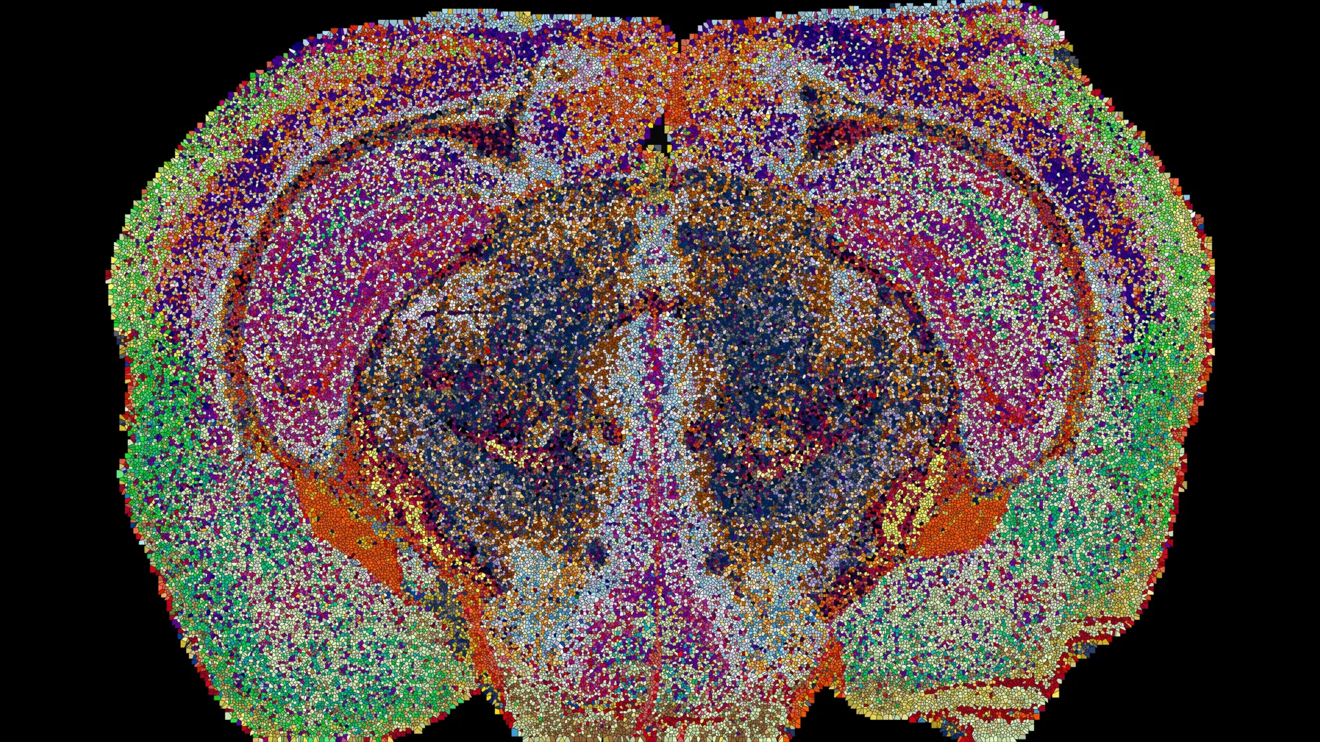
For decades, the mainstream consensus of neurobiology has been dominated by the study of the brain’s cellular architecture, its intricate synaptic connections, and the complex cascades of genes and proteins that dictate neural development. While this gene- and protein-centric approach has yielded monumental breakthroughs in understanding conditions ranging from schizophrenia to neurodegenerative decline, it has historically overlooked an equally vital biological component: lipids. These diverse fatty molecules form the foundational fluid mosaic of the cellular membranes surrounding neurons, construct the essential myelin insulation required for rapid nerve signal propagation, and act as dynamic signaling mediators that facilitate intercellular communication. Yet, despite their fundamental roles in maintaining neurological homeostasis, lipids have remained largely unmapped within the physical landscape of the central nervous system.
Conventional analytical frameworks and neuroimaging techniques have long struggled to resolve the spatial distribution of individual lipid species within intact or sectioned neural tissue. Standard mass spectrometry and biochemical extraction methods often required the homogenization of brain tissue, effectively erasing the micro-spatial architecture and destroying the precise coordinates where these molecules exerted their biochemical functions. Consequently, neuroscientists possessed only a generalized, highly homogenized picture of how lipid profiles varied across distinct anatomical regions. This technical blind spot represented a critical vulnerability in modern neuroscience, particularly as emerging epidemiological and biochemical evidence increasingly implicated lipid dysregulation in a broad spectrum of neuropathology, including major depressive disorder, bipolar disorder, and Alzheimer’s disease.
A collaborative team of researchers at the École Polytechnique Fédérale de Lausanne (EPFL) has now bridged this historical divide. Spearheaded by Professor Giovanni D’Angelo, who holds The Kristian Gerhard Jebsen Foundation Chair in Nutrition and Metabolism, alongside colleagues Luca Fusar Bassini and Gioele La Manno, the research team has successfully constructed a comprehensive, high-resolution 3D map illustrating the precise distribution of lipids throughout the murine brain. Their landmark findings, which redefine how neurobiologists view chemical compartmentalization within the central nervous system, were published in the peer-reviewed journal Nature, instantly establishing a new baseline for molecular neurobiology.
Chronology of Discovery: From Mass Spectrometry to Machine Learning
The creation of the Lipid Brain Atlas was not a sudden breakthrough, but rather the culmination of years of rigorous methodological development, high-throughput experimentation, and advanced computational modeling. The project began as an ambitious attempt to overcome the historical limitations of mass spectrometry imaging (MSI), a powerful analytical technique that allows researchers to detect the spatial localization of specific chemical compounds across thin biological samples. However, scaling MSI to capture the vast and chemically diverse lipidome of an entire mammalian brain presented unprecedented technical hurdles.
In the early stages of the project, the EPFL team focused on optimizing sample preparation and laser desorption parameters to ensure that fragile lipid molecules were not degraded during the scanning process. By utilizing advanced matrix-assisted laser desorption/ionization (MALDI) mass spectrometry imaging, the researchers were able to systematically scan thin slices of brain tissue, one micro-location at a time. Each scan generated a massive cryptographic readout of the specific lipid species present at exact coordinate points within the tissue architecture.
Over successive months of data collection, the team systematically examined 172 distinct lipid species across 109 meticulously prepared brain slices, harvested from a cohort of 11 standardized laboratory mice. This exhaustive analytical pipeline generated an astronomical dataset comprising approximately 7 million individual chemical measurements. Managing, cleaning, and spatially aligning millions of spectral data points required the development of bespoke machine-learning algorithms and computational reconstruction tools. By feeding the mass spectrometry data into these custom algorithms, the research team successfully stitched the two-dimensional tissue slices back into a seamless, high-fidelity three-dimensional digital model of the mouse brain.
Decoding the Lipizones: 539 Distinct Chemical Territories
When the computational reconstruction was finalized, the resulting Lipid Brain Atlas revealed a level of molecular organization that far exceeded initial expectations. Rather than being randomly dispersed or passively distributed, lipid molecules were discovered to be arranged in highly structured, meticulously patterned configurations. Some of these chemical topographies mirrored classic anatomical boundaries long recognized by classical neuroanatomists, while others exposed entirely novel biochemical boundaries that had remained completely invisible to genetic and structural staining techniques.
The data analysis revealed a staggering 539 chemically distinct territories within the mouse brain, which the researchers formally designated as "lipizones." Each lipizone possesses a unique, highly reproducible chemical signature defined by a characteristic stoichiometric combination of specific lipid classes. The distinctiveness of these chemical signatures was so pronounced that automated algorithms could accurately determine the exact anatomical origin of an unknown brain tissue sample solely by analyzing its lipid composition.
Many of these newly identified lipizones aligned with established functional nuclei and distinct cell types. However, a significant subset of the regions unveiled unexpected biochemical relationships. The atlas mapped chemical bridges connecting distant groups of nerve cell bodies with remote axonal projection areas, highlighting functional circuits defined by their shared lipid microenvironments rather than their transcriptional profiles.
"In effect, lipids work like postal codes, giving distant but related parts of the brain the same chemical address," explained Professor D’Angelo during an overview of the findings. This chemical addressing system suggests that lipid distribution is not merely a consequence of cellular structure, but an active, tightly regulated component of neural circuitry and connectivity.
Challenging Established Paradigms in White Matter and Ventricular Lining
The unveiling of the Lipid Brain Atlas has already begun to challenge long-held assumptions regarding the chemical homogeneity of various brain tissues. One of the most striking revelations emerged from the detailed analysis of white matter—the complex neural wiring composed predominantly of myelinated nerve fibers that facilitate rapid communication between disparate cortical and subcortical regions.
Historically, white matter was broadly conceptualized by neuroscientists as a functionally and chemically uniform tract whose primary role was structural insulation and signal conduction. However, the EPFL atlas demonstrated that white matter is partitioned into numerous distinct lipid zones, reflecting a previously unappreciated degree of biochemical heterogeneity.
"This shows that the cells which make myelin are far more varied in their chemistry than their genes alone would suggest," noted Luca Fusar Bassini, highlighting the limitations of relying exclusively on transcriptomics to infer cellular function. oligodendrocytes—the specialized glial cells responsible for synthesizing and maintaining the myelin sheath in the central nervous system—exhibit profound regional metabolic specializations that are directly reflected in their surrounding lipid microenvironments.
In addition to white matter, the research team identified previously unrecognized chemical boundaries within the choroid plexus, the specialized vascular network responsible for producing and regulating cerebrospinal fluid. Similar chemical demarcations were discovered within the ependymal tissues lining the brain’s internal ventricular cavities. Together, these discoveries underscore the profound analytical power of lipid mapping, proving that biochemical organization can reveal critical physiological and structural features of the brain that remain completely obscured when scientists rely solely on classical anatomy or genetic expression profiles.
Pregnancy and Neuroplasticity: Dynamic Shifts in Brain Chemistry
Beyond mapping the steady-state architecture of the healthy adult mouse brain, the EPFL research team deployed the Lipid Brain Atlas to investigate how the brain’s chemical landscape responds to profound physiological challenges, specifically pregnancy. Neurobiologists have long recognized that the maternal brain undergoes extensive functional and structural adaptations to prepare for offspring care, yet the precise molecular mechanisms driving these plastic changes have remained poorly understood.
When the researchers analyzed the brains of pregnant mice using the lipid-mapping pipeline, they observed widespread, systematic shifts in lipid composition across numerous neural regions. Remarkably, the magnitude of these pregnancy-induced lipid fluctuations significantly overshadowed the baseline lipid differences typically observed between non-pregnant male and female mice.
The cerebral cortex, the outermost layer of the brain responsible for higher-order cognitive processing, sensory perception, and motor control, exhibited particularly extensive chemical remodeling. Simultaneously, researchers recorded a sharp, localized surge in the levels of galactosyl ceramide—a critical sphingolipid and essential structural component of myelin—throughout substantial portions of the brain’s white matter tracts.
These findings provide compelling evidence that gestation triggers a massive, coordinated retuning of the central nervous system’s lipidome. This active biochemical remodeling is hypothesized to optimize neural network plasticity, allowing maternal neural circuits to adapt dynamically to the immense physical and behavioral demands of pregnancy and postpartum care.
"Together, these shifts suggest the brain retunes its own wiring to meet the demands of pregnancy," stated Gioele La Manno, emphasizing the adaptive nature of the brain’s lipid metabolism. "The Atlas reveals that the brain constantly adjusts its lipids to meet the body’s changing needs."
Broader Implications, Open Access, and Future Directions
Recognizing the transformative potential of their work for the broader scientific community, the EPFL research team has made the Lipid Brain Atlas entirely open access and freely available online. This interactive digital resource allows neuroscientists, molecular biologists, and clinical researchers worldwide to explore the complex lipid architecture of the mammalian brain without the barrier of specialized mass spectrometry infrastructure.
By integrating this unprecedented spatial lipid data with existing multi-omic repositories—such as single-cell RNA sequencing atlases and high-resolution structural connectomes—researchers can now construct a truly multidimensional model of brain organization. This holistic approach promises to illuminate how localized lipid distributions influence cellular differentiation, synaptic plasticity, and metabolic homeostasis throughout the lifespan.
Furthermore, the atlas provides an invaluable, standardized baseline reference for healthy neural tissue. Investigators can now utilize this reference framework to interrogate how lipid distributions are disrupted during pathological aging, or how specific lipid alterations contribute to the etiology of neurodevelopmental disorders and neurodegenerative conditions such as Alzheimer’s disease and major depression.
While the current atlas is derived exclusively from murine models and cannot be directly translated into a human clinical map without further validation, the underlying analytical methodology offers a powerful roadmap for future human tissue studies. The successful translation of mass spectrometry imaging and machine-learning reconstruction techniques to human post-mortem tissue could soon revolutionize clinical neuropathology.
"What transfers to humans isn’t the map, it’s the method, and the fact that we now know that a healthy brain has a very organized lipid map at all," concluded Luca Fusar Bassini. As the scientific community begins to leverage this new resource, the long-overlooked lipid molecule is finally taking its rightful place at the forefront of modern neurobiological research.
