
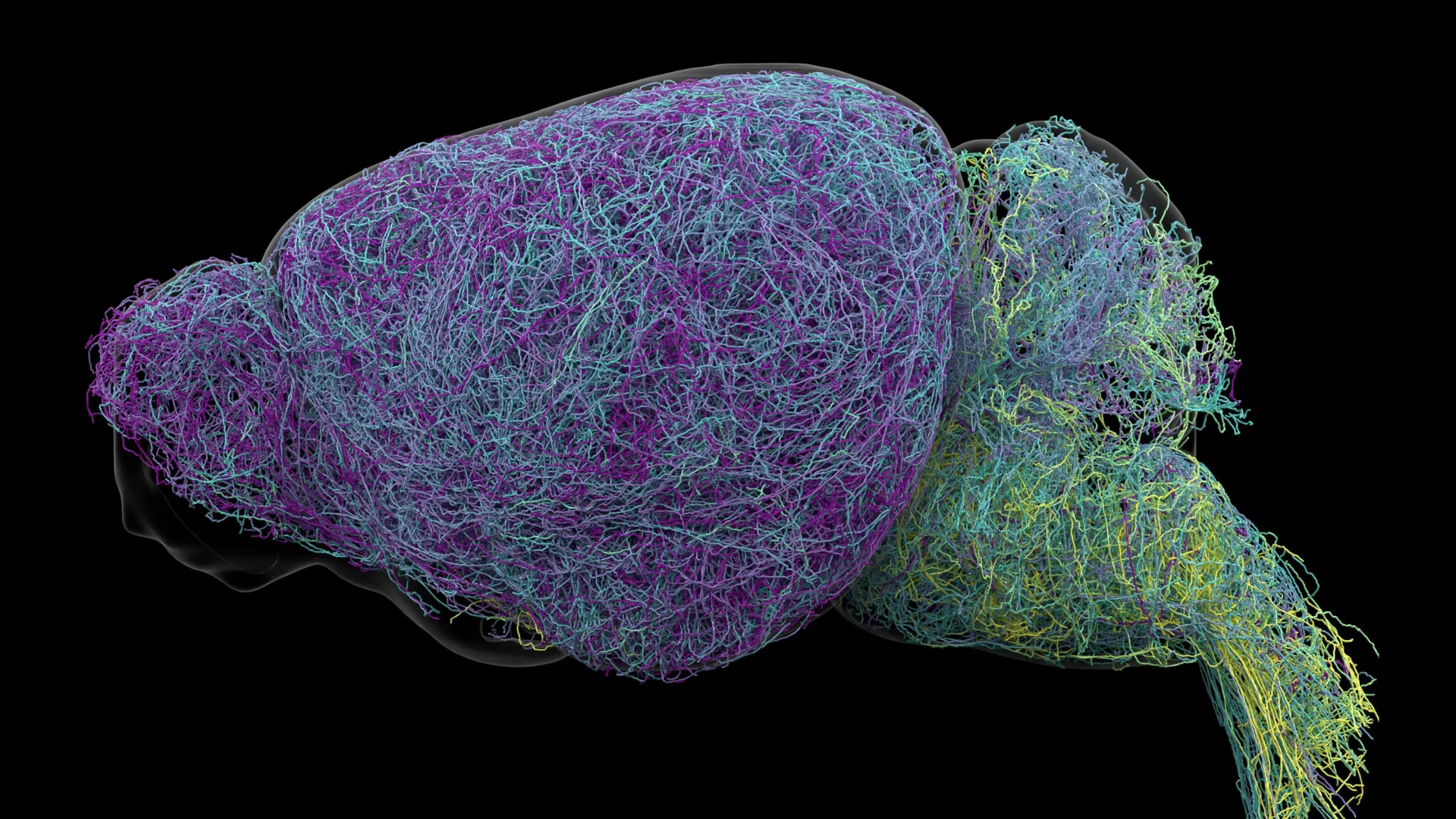
Deep within the brainstem lies a microscopic nucleus that has commanded the attention of neuroscientists for decades: the locus coeruleus, aptly named the "blue place" for its dark pigmentation under a microscope. Despite containing a mere fraction of the total neuronal population within the mammalian central nervous system, this minuscule region exerts an outsized influence over human physiology and behavior. As the primary factory for norepinephrine—a chemical messenger vital for modulating attention, memory formation, arousal, wakefulness, cardiovascular tone, and adaptive responses to environmental stressors—the locus coeruleus has long been characterized by textbooks as the brain’s ultimate broadcasting system. For generations, the prevailing scientific consensus maintained that this structure acted much like a biological loudspeaker, spraying a generalized chemical fog of norepinephrine across vast expanses of the brain indiscriminately.
However, a monumental paradigm shift has arrived from the laboratories of the Allen Institute. In a comprehensive study published in the prestigious journal Nature, an interdisciplinary team of researchers has definitively dismantled the loudspeaker hypothesis. Utilizing a sophisticated arsenal of cutting-edge technologies—including high-resolution whole-brain imaging, real-time electrophysiological recordings of neural activity, high-throughput single-cell transcriptomics, and rigorous behavioral experiments in murine models—the research team revealed that the locus coeruleus does not shout a single, generalized message into the void. Instead, it operates with exquisite precision, communicating through highly segregated, specialized neural pathways that direct distinct chemical signals to specific anatomical destinations based on real-time experiential demands.
This revelation transforms our baseline comprehension of neurochemistry. Rather than a blunt instrument of arousal, the norepinephrine system emerges as a sophisticated, targeted postal network capable of routing precise messages to distinct neural addresses. The implications of this structural and functional mapping extend far beyond basic neurobiology, opening unprecedented avenues for the pharmacological treatment of neuropsychiatric disorders, neurodegenerative diseases, and cognitive deficits.
A Chronological Quest: How Scientists Unraveled the Blueprint of the Blue Place
The journey toward rewriting the neuroanatomy of the locus coeruleus represents the culmination of years of technological innovation and painstaking empirical investigation. For decades, researchers recognized the physiological importance of norepinephrine, largely due to its involvement in the fight-or-flight response and its modulation by widely prescribed medications. Yet, technical limitations prevented scientists from observing the individual projections of locus coeruleus neurons in fine detail. Traditional tracing methods could capture local connectivity or general pathways, but they lacked the resolution necessary to map individual axons across the entire murine brain while simultaneously recording their electrical activity and genetic signatures.
The breakthrough began taking shape as part of larger initiatives funded heavily by the National Institutes of Health through the ambitious Brain Research Through Advancing Innovative Neurotechnologies (BRAIN) Initiative. Over recent years, the Allen Institute assembled a multidisciplinary team tasked with mapping the mammalian brain with unprecedented granularity. By combining multiplexed error-robust fluorescence in situ hybridization (MERFISH), viral tracing techniques, automated serial two-photon tomography, and advanced computational reconstruction software, the researchers set out to construct a comprehensive atlas of the locus coeruleus.
The investigative timeline moved systematically from anatomical mapping to functional correlation. Initially, the team focused on charting the spatial organization of the nucleus. They discovered a clear topographical division: neurons situated in the dorsal, or upper, strata of the locus coeruleus project their axons primarily upward, feeding information into the cerebral cortex and forebrain structures. Conversely, neurons housed within the ventral, or lower, strata direct their axonal outputs downward, communicating primarily with the brainstem and the spinal cord.
Following this structural mapping, the researchers introduced behavioral paradigms to mice navigating decision-making and reward-based tasks. By recording neural activity in real time, the team tracked how dorsal versus ventral populations responded during distinct phases of learning and environmental engagement. The convergence of spatial tracing, genetic profiling of nearly 400,000 individual cells, and behavioral telemetry culminated in the landmark publication in Nature, forever altering the theoretical framework of central nervous system neuromodulation.
Quantitative Brilliance: Inside the Data of a Record-Breaking Neural Network
The scale of the data generated by the Allen Institute study underscores the sheer complexity of the undertaking. To decode the locus coeruleus, the researchers executed a massive quantitative analysis encompassing nearly 35,000 neurons evaluated through whole-brain imaging pipelines, alongside the detailed genetic profiling of approximately 400,000 individual cells.
Among the most astonishing discoveries to emerge from this data torrent was the physical architecture of individual locus coeruleus neurons. Axons—the thread-like projections through which neurons send electrical and chemical impulses to neighboring cells—are typically understood to span local or regional distances depending on their functional class. Within the locus coeruleus, however, the researchers mapped axons of staggering proportions.
On average, the axons projecting from locus coeruleus neurons measured an impressive 35 centimeters in length—a remarkable distance for a mouse brain, where the organ itself spans only a fraction of that scale due to the extensive branching and winding of the projections. More remarkably, the team successfully reconstructed one record-breaking neuron whose total axonal length reached an astonishing 70.32 centimeters, establishing it as the longest single neuron ever meticulously measured and reconstructed in a murine model.
This architectural marvel defies conventional assumptions about biological efficiency. Despite possessing an axon long enough to blanket a massive volume of the cerebral cortex with norepinephrine-releasing terminals, this single neuron exercises extreme selectivity. It deliberately bypasses the cerebellum, brainstem, and spinal cord, focusing its chemical output exclusively on targeted cortical networks. This quantitative finding mathematically disproves the notion of the locus coeruleus as a diffuse foghorn, providing hard empirical evidence of structured, long-range specificity.
Functional Divergence: Decoding Learning and Environmental Engagement
Beyond structural layout and physical dimensions, the study illuminated a profound functional dichotomy between the dorsal and ventral subpopulations of the locus coeruleus, supported by distinct patterns of gene expression.
When observing mice engaged in active learning tasks—specifically scenarios where animals had to reevaluate their choices and adapt their behavior following an unfavorable or unrewarded outcome—neurons in the dorsal portion of the locus coeruleus exhibited dramatic spikes in activity. These cells, projecting upward into the cortex, appear to play a direct mechanical role in cognitive flexibility, error correction, and the assimilation of new experiences into behavioral strategies.
In stark contrast, the ventral population of neurons followed an entirely different behavioral schedule. The electrical firing rates of ventral cells surged shortly before the test subjects chose to ignore available environmental cues or forego potential rewards. This indicates that ventral neurons, which project downward into the brainstem and spinal cord, act as behavioral gatekeepers, modulating the organism’s baseline motivation and regulating whether an animal decides to actively engage with its surroundings or remain quiescent.
Karel Svoboda, director of Neural Dynamics at the Allen Institute and a co-author of the study, encapsulated the finding during a media briefing: "What emerged was a clear map: neurons in the dorsal LC that send signals upward to the cortex are involved in learning, whereas neurons in the ventral region projecting downward to the brainstem and spinal cord govern whether animals engage with their environment at all. We also showed that these anatomical differences are mirrored by distinct gene expression patterns."
These genetic signatures confirm that the functional specialization observed in the laboratory is hardwired into the cellular identity of these neurons, shaped by unique transcriptional profiles that dictate their connectivity, morphology, and physiological roles.
Official Responses and Scientific Implications
The broader scientific and medical communities have received the Allen Institute’s findings with intense enthusiasm, recognizing their potential to reshape pharmacological research. Dr. Jeremiah Cohen, an Allen Institute scientist and co-author of the study, emphasized the uniqueness of the axonal architecture discovered during the project, noting that the capacity of individual cells to maintain target specificity across such vast anatomical distances challenges foundational dogmas in systems neuroscience.
Supported financially by the National Institutes of Health’s BRAIN Initiative—a federal effort designed to accelerate the development and application of innovative technologies to produce a revolutionary dynamic picture of the brain—the research addresses longstanding challenges in clinical pharmacology.
The implications for human health are profound, particularly regarding disorders where norepinephrine dysregulation plays a central pathological role. Conditions such as major depressive disorder, generalized anxiety disorder, and attention deficit hyperactivity disorder (ADHD) are routinely treated with pharmaceutical agents—such as selective norepinephrine reuptake inhibitors (SNRIs) or tricyclic antidepressants—that modulate brainwide norepinephrine signaling. While these medications provide therapeutic relief for many patients, their broad mechanism of action often induces frustrating off-target side effects, such as elevated heart rate, sleep disturbances, or emotional flattening, because they flood multiple, unrelated neural circuits simultaneously.
Furthermore, locus coeruleus neurons are among the very first groups of cells in the human brain to undergo neurodegeneration in the early stages of Alzheimer’s disease, often deteriorating decades before the appearance of hallmark cognitive symptoms or widespread memory loss. Understanding the precise wiring diagram of these vulnerable cells could shed light on why they are selectively susceptible to pathology and how their early loss triggers the downstream cognitive deficits characteristic of dementia.
By mapping the specialized pathways, genetic signatures, and behavioral correlates of the locus coeruleus, modern neuroscience moves one step closer to precision medicine in psychiatry and neurology. Armed with a detailed blueprint of how the brain routes attention and regulates learning, future pharmacologists may be empowered to design next-generation therapeutics capable of selectively targeting individual neural circuits tied to specific symptoms, maximizing clinical efficacy while minimizing collateral disruption to the broader central nervous system.
