
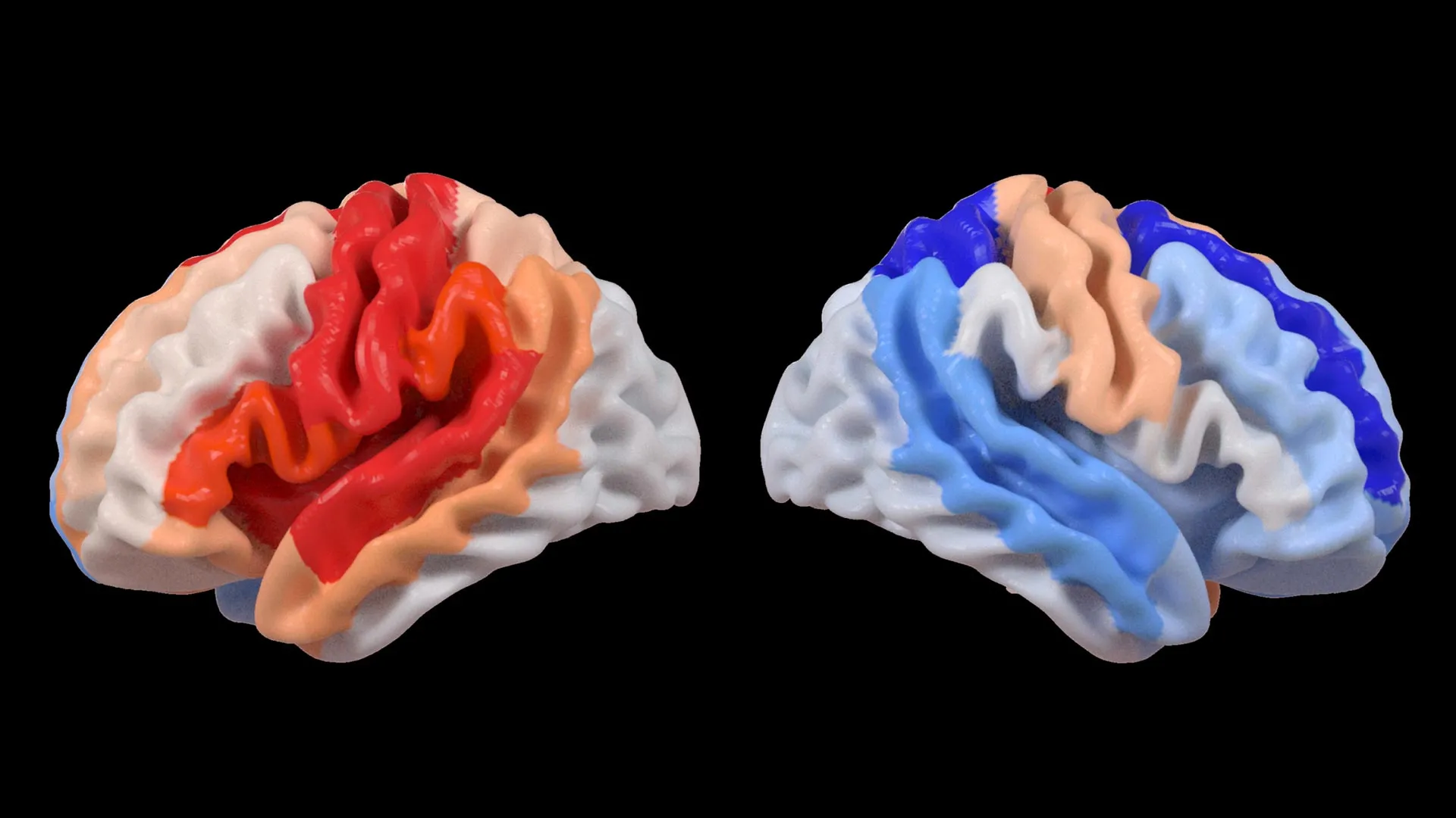
A groundbreaking study published in the esteemed journal The Lancet Digital Health has unveiled a remarkable and previously underappreciated capacity of the human brain to adapt in the wake of a stroke. Researchers at the USC Mark and Mary Stevens Neuroimaging and Informatics Institute (Stevens INI) have identified that individuals exhibiting significant physical disabilities following a stroke may, paradoxically, present with structural characteristics of a "younger" brain in regions that remained unaffected by the initial injury. This phenomenon is believed to be a potent indicator of the brain’s intrinsic ability to reorganize and compensate for lost functionality.
The extensive research was meticulously conducted as a core component of the Enhancing NeuroImaging Genetics through Meta-Analysis (ENIGMA) Stroke Recovery Working Group. This collaborative initiative brought together scientists from across the globe to analyze a substantial dataset comprising brain scans from over 500 stroke survivors. These valuable data were meticulously collected from 34 distinct research centers spanning eight countries, creating an unprecedented global repository of stroke recovery information. To dissect these complex datasets, the team employed sophisticated deep learning models, trained on a vast corpus of tens of thousands of MRI scans. This advanced analytical approach allowed researchers to estimate the "brain age" of specific regions within each hemisphere of the brain, and critically, to examine the intricate interplay between stroke-induced damage, brain structure, and the subsequent trajectory of recovery.
AI Illuminates Brain Rewiring Post-Stroke
At the heart of this groundbreaking investigation lies the application of a cutting-edge artificial intelligence technique known as a graph convolutional network. This advanced AI system was instrumental in estimating the biological age of 18 distinct brain regions based on detailed MRI data. By comparing these AI-generated age estimates with the chronological age of each participant, researchers were able to calculate a metric termed the "brain-predicted age difference" (brain-PAD). This metric serves as a sensitive indicator of overall brain health, with significant deviations from expected age potentially signaling underlying pathology or compensatory mechanisms.
The research team then meticulously correlated these brain age measurements with standardized scores assessing motor function. A strikingly clear pattern emerged: stroke survivors who experienced severe impairments in movement, even after more than six months of dedicated rehabilitation, exhibited a younger-than-expected brain age in brain regions situated on the side opposite to the stroke’s origin. This effect was particularly pronounced within the frontoparietal network, a critical brain circuit known to be involved in complex cognitive functions such as movement planning, sustained attention, and motor coordination.
"We were astonished to observe that while larger strokes tend to accelerate the aging process in the damaged hemisphere, the opposite hemisphere paradoxically appears younger," stated Hosung Kim, PhD, an associate professor of research neurology at the Keck School of Medicine of USC and a co-senior author of the study. "This distinct pattern strongly suggests that the brain is actively engaged in a process of profound reorganization, essentially rejuvenating undamaged neural networks to compensate for the functional deficits caused by the stroke."
Dr. Kim further elaborated on the significance of these findings: "These results indicate that when stroke damage leads to a greater loss of motor control, undamaged regions on the contralateral side of the brain may adapt and actively contribute to functional compensation. We specifically observed this phenomenon in the contralesional frontoparietal network, which displayed a more ‘youthful’ structural pattern. This network is well-established to support motor planning, attention, and coordination. Rather than solely indicating a complete restoration of movement, this observed pattern may represent the brain’s sophisticated attempt to adjust and adapt when the primary motor system is compromised and can no longer operate at its full capacity. This discovery provides us with an entirely new lens through which to observe neuroplasticity, a level of detail that traditional neuroimaging techniques have previously been unable to capture."
Leveraging Global Data for Unveiling Hidden Neural Patterns
The success of this monumental study hinges significantly on the ENIGMA initiative, a far-reaching global collaboration that aggregates and standardizes brain imaging and clinical data from over 50 countries. This unprecedented pooling of resources aims to foster a deeper and more comprehensive understanding of the human brain across a diverse spectrum of neurological conditions. By harmonizing MRI data and clinical information from numerous independent research groups, the team successfully constructed what is believed to be the largest and most comprehensive stroke neuroimaging dataset of its kind ever assembled.
"By pooling data from hundreds of stroke survivors worldwide and harnessing the power of cutting-edge artificial intelligence, we are now capable of detecting incredibly subtle patterns of brain reorganization that would remain entirely invisible in smaller, more localized studies," emphasized Arthur W. Toga, PhD, director of the Stevens INI and Provost Professor at USC. "These findings, revealing regionally differential brain aging in individuals with chronic stroke, hold the profound potential to guide the development of highly personalized and effective rehabilitation strategies in the future."
A New Horizon for Personalized Stroke Recovery
The research team is not resting on their laurels. Their immediate future plans involve longitudinal tracking of patients, observing their brain structure and functional recovery from the acute phase immediately following a stroke through to the long-term recovery period. By meticulously charting how brain aging patterns and structural modifications evolve over time, clinicians and researchers anticipate being able to tailor rehabilitation interventions to each individual’s unique recovery trajectory. The ultimate goal of this ongoing research is to significantly improve functional outcomes and enhance the overall quality of life for stroke survivors.
Background and Context
Stroke, a sudden interruption of blood supply to the brain, remains a leading cause of long-term disability worldwide. According to the World Health Organization, an estimated 15 million people suffer a stroke each year. The impact of a stroke extends far beyond immediate survival, often resulting in devastating physical, cognitive, and emotional challenges for survivors and their families. Motor impairments, such as paralysis or weakness on one side of the body, are among the most common and debilitating consequences, significantly impacting independence and daily functioning.
Historically, stroke recovery research has focused on understanding the mechanisms of brain damage and identifying therapeutic interventions to promote neuronal repair. Neuroplasticity, the brain’s remarkable ability to reorganize itself by forming new neural connections throughout life, has long been recognized as a critical factor in functional recovery. However, the precise ways in which neuroplasticity manifests, particularly in the context of chronic stroke and severe motor deficits, have remained elusive.
The advent of advanced neuroimaging techniques, such as Magnetic Resonance Imaging (MRI), has provided unprecedented insights into brain structure and function. However, analyzing the subtle changes associated with neuroplasticity, especially in large, diverse populations, presents significant computational and analytical challenges. This is where the power of artificial intelligence, particularly deep learning, has emerged as a transformative tool.
Timeline of the Study
While the publication date marks the culmination of this research, the journey began with the establishment and ongoing work of the ENIGMA Stroke Recovery Working Group. This collaborative effort likely involved years of data acquisition, standardization, and preliminary analysis across participating research institutions. The deep learning models used in the study would have required extensive training periods, drawing upon vast libraries of MRI scans beyond the specific stroke survivor cohort. The analysis of the 500+ stroke survivor scans and their correlation with clinical motor function scores represents a significant phase of dedicated scientific investigation. The findings, presented in The Lancet Digital Health, represent the latest milestone in this ongoing global effort to unravel the complexities of stroke recovery.
Supporting Data and Implications
The study’s strength lies in its unprecedented scale. Analyzing data from over 500 stroke survivors across 34 centers in eight countries provides a robust statistical foundation, reducing the likelihood of findings being due to chance or specific local factors. The use of deep learning, a sophisticated form of AI, allowed for the detection of patterns that are simply not discernible through traditional visual inspection of MRI scans or simpler analytical methods.
The concept of "brain age" is a powerful metric. While a damaged hemisphere might exhibit signs of accelerated aging due to cellular stress and neuronal loss, the observed "youthfulness" in the contralateral hemisphere suggests a proactive and adaptive response. This phenomenon is particularly intriguing in the context of the frontoparietal network. This network is crucial for higher-level cognitive functions that are intrinsically linked to motor control, such as planning a movement, directing attention to relevant stimuli, and coordinating complex motor sequences. The finding that this network appears "younger" in individuals with severe motor impairments suggests it is being actively recruited and potentially "rejuvenated" to try and compensate for the lost function in the damaged motor pathways.
The implications of this research are far-reaching:
- Understanding Neuroplasticity: This study offers a novel and quantitative way to observe neuroplasticity in action. It moves beyond simply stating that the brain is plastic to demonstrating how specific brain regions adapt and potentially "rejuvenate" to compensate for injury.
- Personalized Rehabilitation: By identifying these compensatory patterns, clinicians may be able to develop more targeted rehabilitation strategies. For example, therapies could be designed to further stimulate and strengthen these "younger," contralesional networks, thereby enhancing their compensatory capabilities.
- Biomarker Development: The brain-PAD metric, derived from regional brain age estimation, could potentially serve as a biomarker for predicting recovery potential or monitoring the effectiveness of interventions.
- Future Research Directions: The study opens new avenues for research into the molecular and cellular mechanisms underlying this observed rejuvenation and compensatory plasticity. Understanding these mechanisms could lead to novel therapeutic targets.
Official and Expert Reactions (Inferred)
While direct quotes from external parties are not provided in the original content, the significance of this publication in The Lancet Digital Health, a highly respected medical journal, suggests strong endorsement from the scientific community. Journals of this caliber undergo rigorous peer review, meaning that independent experts in the field have scrutinized and validated the methodology and findings.
It can be reasonably inferred that stroke rehabilitation specialists and neurologists will view these findings with considerable interest. They represent a tangible step forward in understanding how the brain copes with devastating injury and offer a glimmer of hope for improving outcomes for patients with severe motor deficits. Neuroscientists involved in AI research will likely be encouraged by the demonstration of deep learning’s power in uncovering complex biological phenomena.
Broader Impact and Future Directions
The ENIGMA initiative itself represents a significant shift in how large-scale neuroscience research is conducted, emphasizing global collaboration and data sharing. This study is a testament to the power of such collaborative efforts in tackling complex scientific questions that are beyond the scope of single institutions.
The researchers’ stated intention to conduct longitudinal studies is crucial. Tracking these brain aging patterns over time will provide invaluable information on the dynamics of neuroplasticity. It will help answer questions such as: Is this "youthfulness" a transient response, or does it persist and contribute to long-term functional gains? Can interventions accelerate or enhance this process?
Ultimately, the goal of improving outcomes and quality of life for stroke survivors is paramount. By providing a deeper understanding of the brain’s remarkable adaptive capabilities, this research paves the way for more effective, personalized, and potentially transformative approaches to stroke rehabilitation. The integration of AI into neuroimaging analysis is not just a technological advancement; it is a paradigm shift that is unlocking new possibilities for understanding and treating complex neurological conditions.
The study, titled "Deep learning prediction of MRI-based regional brain age reveals contralesional neuroplasticity associated with severe motor impairment in chronic stroke: A worldwide ENIGMA study," received funding from the National Institutes of Health (NIH) under grant R01 NS115845. The research was further supported by international collaborators from institutions including the University of British Columbia, Monash University, Emory University, and the University of Oslo, underscoring the truly global nature of this scientific endeavor. Further insights into the associations between contralesional neuroplasticity and motor impairment can be gained by viewing an explanatory video produced by the Stevens INI.
