Researchers at Johns Hopkins Medicine, bolstered by significant funding from the National Institutes of Health (NIH), are pioneering a groundbreaking approach to Alzheimer’s disease treatment, focusing on a crucial protein in the brain responsible for producing a seemingly unassuming yet vital gas. This innovative research delves into the intricate mechanisms of memory formation and offers a new perspective on neuroprotection, potentially paving the way for novel therapeutic interventions against this devastating neurodegenerative condition that affects millions worldwide.
The Pivotal Role of Cystathionine γ-lyase and Hydrogen Sulfide in Cognitive Function
At the heart of this research lies the protein Cystathionine γ-lyase, or CSE. While best known for its role in generating hydrogen sulfide (H₂S) – a gas infamous for its pungent, rotten-egg odor – CSE is now understood to play a far more significant and nuanced role in the brain, particularly in the complex processes that underpin memory formation and cognitive health. This revelation stems from extensive experiments conducted on genetically engineered mice, spearheaded by Dr. Bindu Paul, an associate professor of pharmacology, psychiatry, and neuroscience at the Johns Hopkins University School of Medicine.
The findings, meticulously detailed in a recent publication in the prestigious journal Proceedings of the National Academy of Sciences, aim to unravel the precise workings of CSE and explore whether enhancing its activity could offer a protective shield for brain cells, thereby slowing the relentless progression of neurodegenerative diseases like Alzheimer’s. This line of inquiry represents a significant leap forward from earlier understandings, moving beyond the known toxicity of H₂S to explore its delicate and beneficial physiological roles at minuscule concentrations.
Hydrogen Sulfide: A Double-Edged Sword in Neuroprotection
The potential neuroprotective properties of hydrogen sulfide have been a subject of scientific intrigue for some time. Previous studies had indicated that H₂S could indeed offer a degree of protection to neurons in animal models. However, a significant hurdle emerged: the inherent toxicity of H₂S when present in larger quantities, rendering direct administration to the brain an unsafe proposition. This inherent challenge necessitated a shift in scientific strategy, compelling researchers to investigate how to safely maintain the exceptionally low levels of H₂S that are naturally and beneficially present within neuronal environments.
The latest research from Johns Hopkins provides compelling evidence supporting this focus. Mice genetically engineered to be deficient in the CSE enzyme exhibited marked impairments in memory and learning capabilities. Crucially, these mice also displayed hallmarks commonly associated with Alzheimer’s disease, including elevated levels of oxidative stress, increased DNA damage, and compromised integrity of the blood-brain barrier. Dr. Paul, the study’s corresponding author, emphasized that these findings strongly suggest a direct link between CSE deficiency and the pathological changes observed in neurodegenerative conditions.
A Legacy of Research: Building Towards a Therapeutic Breakthrough
This current body of work stands on the shoulders of decades of foundational research, particularly the pioneering investigations led by Dr. Solomon Snyder, a professor emeritus of neuroscience, pharmacology, and psychiatry at Johns Hopkins. Dr. Snyder’s lab has been instrumental in uncovering the physiological roles of various signaling molecules in the brain.
A pivotal moment in this research trajectory occurred in 2014 when Dr. Snyder’s team reported that CSE played a supportive role in brain health, specifically in mouse models of Huntington’s disease. This earlier work utilized mice lacking the CSE protein, a strain initially developed in 2008 when the protein’s connection to vascular function and blood pressure regulation was first elucidated. These early investigations laid the groundwork for understanding CSE’s broader impact on neurological health.
Further building on this foundation, in 2021, the research group observed that CSE function was impaired in mouse models of Alzheimer’s disease. In a promising development, very small, carefully controlled injections of hydrogen sulfide were found to help protect brain function in these models. While these earlier studies often involved mice with additional genetic mutations predisposing them to neurodegenerative diseases, the latest research has the distinct advantage of isolating and focusing solely on the role of CSE itself, providing a clearer understanding of its independent contribution to cognitive health.
Dr. Snyder, who retired from the Johns Hopkins Medicine faculty in 2023 but remains a co-corresponding author on the current study, articulated the significance of these latest findings: "This most recent work indicates that CSE alone is a major player in cognitive function and could provide a new avenue for treatment pathways in Alzheimer’s disease." This statement underscores the potential of CSE as a novel therapeutic target, moving beyond a symptomatic approach to addressing the underlying biological mechanisms of the disease.
Chronology of Key Discoveries in CSE and Neurodegeneration Research:
- 2008: Development of mice lacking the CSE protein, initially linked to blood vessel function and blood pressure regulation.
- 2014: Dr. Snyder’s team reports CSE’s role in supporting brain health in Huntington’s disease mouse models.
- 2021: Research demonstrates impaired CSE function in Alzheimer’s disease mouse models, with H₂S injections showing protective effects.
- Present: The current study isolates CSE’s role in cognitive function, revealing its critical impact on memory and neuroprotection, independent of other genetic factors.
Memory Deficits Directly Correlated with CSE Deficiency
To meticulously investigate the link between CSE and memory, the research team employed a comparative approach, contrasting mice lacking the CSE protein with their genetically normal counterparts from the established 2008 strain. A key experimental tool utilized was the Barnes maze, a widely recognized test designed to assess spatial memory – the ability to recall directions and navigate environments.
In this test, mice are trained to locate a hidden shelter to escape a bright, aversive light. At two months of age, both the normal mice and those lacking CSE demonstrated comparable performance, successfully finding the shelter within a three-minute timeframe. However, a significant divergence emerged by the six-month mark. The CSE-deficient mice began to struggle considerably in locating the escape route, indicating a progressive decline in their spatial memory. In stark contrast, the normal mice continued to perform with consistent success.
"The decline in spatial memory indicates a progressive onset of neurodegenerative disease that we can attribute to CSE loss," stated Dr. Suwarna Chakraborty, the study’s first author and a researcher in Dr. Paul’s laboratory. This observation provides concrete behavioral evidence linking the absence of CSE to impaired cognitive function, a hallmark of Alzheimer’s disease.
Behavioral Evidence: Barnes Maze Performance Over Time
| Mouse Group | Age (Months) | Average Time to Find Shelter (Minutes) | Observations |
|---|---|---|---|
| Normal (CSE Present) | 2 | < 3 | Efficient navigation and shelter location. |
| CSE-Deficient | 2 | < 3 | Comparable performance to normal mice, indicating early-stage normalcy. |
| Normal (CSE Present) | 6 | < 3 | Continued successful navigation and shelter location. |
| CSE-Deficient | 6 | > 3 (significant increase) | Marked difficulty in finding the escape route, indicating progressive memory loss. |
This data clearly illustrates the detrimental impact of CSE deficiency on learning and memory retention as the mice age.
Cellular and Structural Brain Changes Mimicking Alzheimer’s Pathology
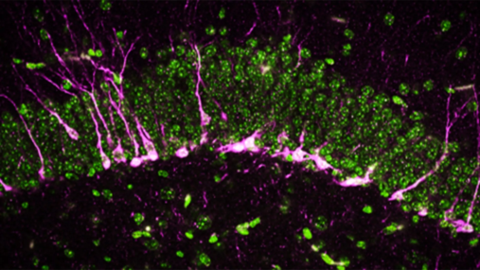
Beyond behavioral observations, the researchers delved into the cellular and structural alterations occurring within the brains of mice lacking CSE. The hippocampus, a brain region critically involved in the consolidation of new memories and spatial navigation, relies heavily on the continuous process of neurogenesis – the formation of new neurons. Disruptions in this delicate process are a well-established characteristic of various neurodegenerative diseases, including Alzheimer’s.
Employing sophisticated biochemical and analytical techniques, the research team identified a significant reduction or complete absence of proteins essential for neurogenesis in the brains of CSE-deficient mice. This finding suggests that the absence of CSE directly interferes with the brain’s ability to generate new, functional neurons.
Further microscopic examination using high-powered electron microscopes revealed profound structural damage within the brains of these mice. Notably, large breaks were observed in blood vessels, indicating a significant compromise of the blood-brain barrier. This breakdown is a critical and well-documented feature of Alzheimer’s disease, allowing harmful substances to enter the brain and exacerbating neuronal damage. Moreover, the study observed that newly formed neurons in these mice encountered difficulties in migrating to the hippocampus, where their integration is vital for memory formation.
Dr. Sunil Jamuna Tripathi, a co-first author and researcher in Dr. Paul’s lab, summarized these cellular findings: "The mice lacking CSE were compromised at multiple levels, which correlated with symptoms that we see in Alzheimer’s disease." This multi-faceted evidence paints a clear picture of how CSE deficiency translates into a cascade of pathological events mirroring those seen in human Alzheimer’s patients.
Key Brain Changes Observed in CSE-Deficient Mice:
- Reduced Neurogenesis: Impaired formation of new neurons, particularly in the hippocampus.
- Blood-Brain Barrier Dysfunction: Evidence of breaks in blood vessels, allowing detrimental substances into the brain.
- Disrupted Neuronal Migration: Newly formed neurons struggle to reach their intended destinations within the brain.
- Increased Oxidative Stress and DNA Damage: Cellular markers indicative of damage and aging.
Implications for Alzheimer’s Disease Treatment: A New Therapeutic Frontier
Alzheimer’s disease represents a formidable public health challenge, affecting an estimated over 6 million individuals in the United States alone, with projections indicating a continued rise in prevalence. Despite decades of research, the current therapeutic landscape offers limited options, with no consistently proven treatments capable of halting or significantly slowing the disease’s relentless progression. The emotional and financial toll on patients, families, and healthcare systems is immense.
The findings from Johns Hopkins Medicine offer a beacon of hope by identifying a novel therapeutic target. The researchers propose that specifically targeting CSE and modulating its production of hydrogen sulfide could unlock a new paradigm for developing therapies. Such therapies would aim not merely to manage symptoms but to protect fundamental brain functions and potentially slow the underlying disease processes. This could involve developing strategies to enhance CSE activity or find ways to safely deliver or mimic the beneficial effects of endogenous H₂S at precise physiological levels.
The substantial funding secured from the National Institutes of Health, alongside contributions from numerous other esteemed organizations, underscores the recognized potential of this research. This multidisciplinary support highlights a collective commitment to advancing our understanding of neurodegenerative diseases and translating scientific discoveries into tangible clinical benefits.
Funding and Collaborative Research Efforts:
The comprehensive scope of this research is supported by a broad coalition of funding bodies, including:
- National Institutes of Health (NIH): Providing significant grants (e.g., 1R01AG071512, P50 DA044123, 1R21AG073684, O1AGs066707, U01 AG073323, AG077396, NS101967, NS133688, P01CA236778).
- Department of Defense: Offering support for critical research initiatives (HT94252310443).
- American Heart Association: Including specific funding through the AHA-Allen Initiative in Brain Health and Cognitive Impairment.
- Solve ME/CFS Initiative.
- Johns Hopkins University Catalyst Award.
- Valour Foundation and Wick Foundation.
- Department of Veterans Affairs Merit Award (I01BX005976).
- Louis Stokes Cleveland Department of Medical Affairs Veterans Center.
- Mary Alice Smith Funds for Neuropsychiatry Research.
- Lincoln Neurotherapeutics Research Fund.
- Gordon and Evie Safran Neuropsychiatry Fund.
- Leonard Krieger Fund of the Cleveland Foundation.
This extensive network of support highlights the collaborative spirit and the perceived importance of this research trajectory within the scientific community. The contributions extend beyond funding, with a diverse team of researchers from various institutions, including Case Western University, Leibniz Institute for Analytical Sciences (Germany), Hollings Cancer Center, Darby Children’s Research Institute, Medical University of South Carolina, and West Virginia University School of Medicine, all playing vital roles in the success of these investigations. This collaborative approach is essential for tackling complex diseases like Alzheimer’s and accelerating the pace of discovery.
The path from laboratory discovery to clinical application is often long and arduous, but the identification of CSE and its role in H₂S production offers a tangible and promising new avenue in the fight against Alzheimer’s disease. Continued research and dedicated investment in this area hold the potential to bring much-needed relief and improved outcomes to millions affected by this devastating condition.
