
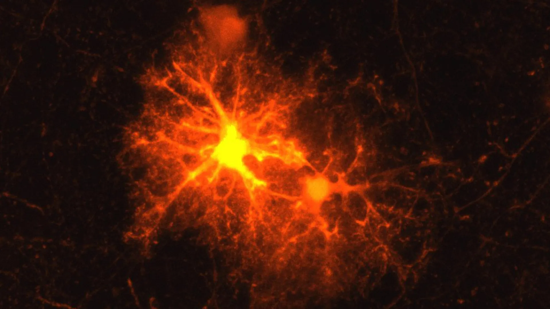
The human brain is often heralded as the most complex structure in the known universe, a delicate network of roughly eighty-six billion neurons operating in a perpetual symphony of electrical and chemical signals. Yet, for all its astonishing computational power, the central nervous system has historically struggled with a notorious vulnerability: when physical damage or neurodegenerative disease strikes, its capacity for self-repair is severely restricted. For decades, neurobiologists have grappled with the mechanisms of neural injury, searching for the cellular triggers that govern degeneration and recovery. Now, a groundbreaking study conducted at the Max Planck Institute of Psychiatry in Munich, Germany, has unveiled a startling and unexpected participant in the brain’s internal emergency response: a classic stress hormone operating within a class of unsung neural repair cells.
For experienced neurobiologist Dr. Jan Deussing, a research group leader at the Max Planck Institute, the phenomenon had long been a familiar visual puzzle. Whenever laboratory mice experienced localized brain damage—such as a precision injection causing minor trauma—Deussing repeatedly observed an identical cellular reaction. A distinct, highly active population of cells would rapidly congregate and proliferate directly around the periphery of the injury site. Although Deussing had witnessed this specialized accumulation countless times over the course of his career, the precise identity of these responding cells and their exact functional role in the broader healing cascade remained an elusive scientific mystery.
A Master Student Takes on a Cellular Enigma
This persistent neurobiological question ultimately transformed into an ideal project for an ambitious master’s student. Clemens Ries, nearing the completion of his biology degree, joined the Max Planck Institute of Psychiatry for a practical internship. Tasked with cracking the identity of the mysterious injury-zone cells, Ries embarked on a systematic investigation that would eventually define the trajectory of his early scientific career.
Working with a standard murine (mouse) model of localized brain injury, Ries employed a methodical immunohistochemical approach. He systematically screened cellular markers corresponding to every known constituent cell type within the central nervous system. After rigorous testing, only a single molecular marker yielded a positive response: the hallmark identifier for oligodendrocyte progenitor cells, commonly known as OPCs.
To fully grasp the magnitude of this discovery, one must understand the critical physiological architecture of the brain. OPCs are specialized precursor cells whose primary developmental destiny is to mature into oligodendrocytes. These specialized glial cells are responsible for generating the myelin sheath—a rich, fatty, multi-layered membrane that wraps around the axons of neurons. Axons serve as the long-distance transmission cables of the nervous system, allowing disparate regions of the brain to communicate at high velocities. Myelin functions analogously to the synthetic insulation wrapped around an electrical wire. Beyond merely accelerating the transmission of electrical impulses, myelin provides essential metabolic and nutritional support to the underlying axons, making it an absolute prerequisite for healthy, stable neurological function.
When myelin is compromised, the consequences are catastrophic. In autoimmune pathologies such as multiple sclerosis (MS), the body’s immune system mistakenly attacks and degrades the protective myelin coating, leading to progressive neurological disability. Physical trauma, ischemia, and neurodegenerative disorders can similarly strip axons of their insulating sheath. In severe cases, uninsulated axons degenerate, culminating in irreversible neuronal cell death. Consequently, the rapid and effective restoration of myelin around damaged axons—a process termed remyelination—is arguably the single most critical component of the brain’s recovery program following injury.
Recognizing the profound implications of his initial findings, Ries transitioned his master’s thesis into a comprehensive doctoral research project under the mentorship of Deussing. What followed was a deep dive into the behavior of OPCs in injured neural tissue, culminating in a series of discoveries recently published in the esteemed scientific journal Cell Reports.
The Injury Response: A Rapid Burst of Stress Hormones
As Ries expanded his investigation, he tracked the behavior of OPCs surrounding brain wounds in real-time. His subsequent data revealed that these progenitor cells undergo a dramatic burst of proliferation at the margins of the lesion. The vast majority of these newly minted cells continue along their developmental trajectory, successfully maturing into functional oligodendrocytes capable of laying down fresh myelin to cover exposed axons.
However, the researchers also stumbled upon a completely unprecedented biochemical twist. In the immediate vicinity of the damaged neural tissue, approximately one-third of the reactive OPCs begin actively transcribing and releasing corticotropin-releasing hormone (CRH). Historically, CRH has been understood by endocrinologists and neuroscientists as a master regulator of the hypothalamic-pituitary-adrenal (HPA) axis—the body’s central command system for managing stress. Released by the hypothalamus, CRH triggers a cascade of hormonal events that prepare the body for "fight or flight."
Prior to this study, the scientific community operated under the rigid assumption that CRH production was largely the domain of specialized neuroendocrine cells and neurons, rather than glial progenitor populations. The revelation that OPCs can synthesize and secrete neuropeptides like CRH fundamentally reshapes our understanding of intercellular communication within injured brain tissue.
The timeline of this CRH release is remarkably precise. Advanced analytical techniques revealed that CRH production by OPCs initiates within mere hours of the initial brain trauma. Yet, this localized endocrine surge is remarkably short-lived; production tapers off and ceases entirely roughly three days post-injury. This narrow, high-intensity window of hormone activity strongly implies that CRH plays a vital signaling role specifically tailored to the hyper-acute phase of the wound-healing response.
Fine-Tuning the Timeline: How CRH Regulates Repair
To determine the functional purpose of this injury-induced CRH burst, Ries and his colleagues investigated how surrounding cells detect and interpret the hormone. The brain utilizes specific membrane-bound proteins to bind CRH, principal among them being corticotropin-releasing hormone receptor 1 (CRHR1).
The team discovered that CRHR1 is densely expressed on a separate, distinct population of OPCs within the local microenvironment. This receptor acts as a cellular antenna, allowing these neighboring progenitor cells to intercept and respond to the CRH secreted by their newly activated counterparts.
To test the physiological necessity of this receptor signaling, the researchers evaluated transgenic mouse models genetically engineered to lack CRHR1. The results of these knockout experiments were striking. In the absence of CRHR1 signaling, injured OPCs initially multiplied at an even faster rate than normal, creating a larger initial pool of precursor cells. However, this early quantitative surge was entirely deceptive. The accelerated proliferation did not translate into enhanced tissue repair. Instead, the maturation process stalled; ultimately, fewer mature oligodendrocytes successfully differentiated, and long-term remyelination was severely impaired.
These experimental insights indicate that CRH does not merely stimulate cell division; rather, it acts as a critical temporal regulator. By modulating the rate at which OPCs transition from proliferation to differentiation, CRH ensures that the regenerative response is properly paced, yielding a sufficient population of mature oligodendrocytes capable of fully restoring the damaged myelin sheath.
From Injury to Development: Shaping the Healthy Brain
Intrigued by the omnipresence of CRHR1 on OPCs, the research team began to ponder a broader biological question. OPCs are not merely dormant reserve cells waiting for a catastrophic injury to occur; they are profoundly active throughout standard brain development. In both mice and humans, the vast majority of myelination takes place postnatally, continuing dynamically through adolescence and into young adulthood as neural networks solidify.
Given that CRHR1 is expressed on OPCs even in the complete absence of physical trauma, Deussing and Ries hypothesized that the exact same receptor-hormone axis might govern normal, developmental myelination. Collaborating with specialized imaging and histology teams, the researchers examined brain tissue architecture in developmental mouse models lacking CRHR1.
The structural analysis confirmed their suspicions. Mice deficient in CRHR1 exhibited systemic alterations in OPC dynamics during early development. Specifically, they produced an abnormally high number of progenitor cells during embryonic and early postnatal growth stages. Crucially, these developmental anomalies were not transient; they permanently altered the neuro-anatomical architecture of the adult brain.
When examining adult brains lacking the receptor, the researchers observed pronounced, permanent hyper-myelination. Detailed cellular tracing revealed that this surplus of myelin was primarily driven by disproportionately thick myelin sheaths wrapping around thin axons. This discovery bridges a major conceptual gap in neurobiology, demonstrating that the CRH-CRHR1 signaling network is a dual-purpose master switch—vital both for orchestrating emergency repairs after trauma and for fine-tuning the baseline structural maturation of the healthy brain.
Unanswered Questions: The Source of Developmental CRH
While the post-injury source of CRH is clearly the reactive OPCs themselves, the discovery of CRH’s role in normal brain development introduces a compelling biological mystery: where does the stress hormone originate when the brain is developing under normal, injury-free conditions?
While definitive proof remains the subject of ongoing investigation, the Max Planck team has formulated a well-supported physiological hypothesis. They propose that developing neurons serve as the primary endogenous source of CRH during ontogeny. According to this model, healthy neurons release baseline amounts of CRH into the extracellular matrix during active developmental windows. This neuronal signal is then captured by CRHR1 on neighboring OPCs, dictating the pace at which progenitor cells multiply and subsequently mature into myelin-producing oligodendrocytes.
This neuron-glia signaling axis highlights a profound level of cooperative crosstalk within the central nervous system, where neuroendocrine stress factors double as essential developmental trophic agents.
Implications for Mental Health and Stress-Associated Disorders
Perhaps the most far-reaching and provocative implication of this research extends beyond regenerative medicine and directly into the realm of psychiatry. It has long been established that neurons release CRH in response to psychological stress, emotional trauma, and environmental adversity. Furthermore, clinical epidemiology consistently identifies early-life stress as a major environmental risk factor for the development of psychiatric disorders later in life, including major depressive disorder, anxiety, and post-traumatic stress disorder (PTSD).
By establishing that OPCs and the myelin-forming apparatus are direct targets of CRH signaling, the Max Planck study introduces a novel biological mechanism linking stress physiology directly to structural brain plasticity.
"Our current findings suggest that in stress-associated psychiatric disorders such as depression, the CRH system in OPCs may play a greater role than previously known," Dr. Deussing speculates.
If subsequent clinical and translational research validates this hypothesized connection, the implications could be transformative. Modern psychiatric pharmacology has long focused primarily on neuronal neurotransmitter systems, such as serotonin, dopamine, and norepinephrine. However, if chronic stress deregulates CRH-mediated OPC maturation and structural myelination, future therapeutic interventions might target glial cells and myelin regeneration pathways directly.
As Clemens Ries transitions from his foundational master’s work to leading a broader program of investigative neurobiology, the scientific community awaits follow-up studies designed to test these psychiatric hypotheses. Unlocking the hidden language of stress hormones within the brain’s repair cells may ultimately provide neurologists and psychiatrists with an entirely unprecedented toolkit for healing the injured and diseased mind.
