
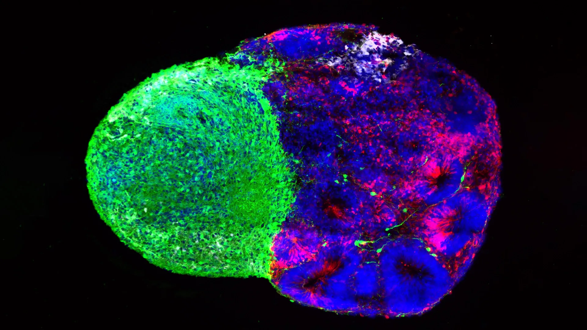
Long before a human infant draws its first breath, an intricate and monumental biological construction project takes place inside the womb. Billions of cellular decisions occur in rapid succession, laying down the neural foundations for thought, emotion, memory, and language. At the very center of this complex orchestration are radial glia—a specialized class of neural stem cells that serve as the primary architects of the human brain. These remarkable cells are largely responsible for generating the vast cellular diversity of the cerebral cortex, the outer layer of the brain that sets humans apart from other species through its unprecedented size and complexity.
Despite their critical importance in prenatal development, radial glia have long remained elusive to scientists. Most of these cells disappear naturally before birth, yet they can mysteriously reemerge decades later within malignant brain tumors, driving aggressive growth through mechanisms that researchers are only beginning to decipher. Now, two groundbreaking studies published simultaneously in the prestigious journals Cell and Science have pierced the veil of early human neurodevelopment. Led by a team of researchers at the University of California, Los Angeles (UCLA), the new research reveals that radial glia do not develop in isolation. Instead, their cellular fates are actively sculpted by two distinct forces: internal metabolic processing and direct physical signaling from distant regions of the developing brain.
The findings not only rewrite foundational textbooks regarding how the cerebral cortex achieves its remarkable variety of cell types, but they also offer unprecedented windows into neurodevelopmental and neuropsychiatric disorders, including autism spectrum disorder, as well as the origins of brain cancer.
The Master Architects: Understanding Radial Glia
To appreciate the significance of the UCLA findings, neuroscientists emphasize the unique evolutionary trajectory of the human brain. Compared with other mammalian species, particularly rodents commonly used in laboratory research, the human cerebral cortex has undergone a massive expansion. This expansion enables advanced cognitive functions such as abstract reasoning, complex language processing, and episodic memory.
Radial glia are the driving force behind this evolutionary leap. Acting as primary neural stem cells, they produce vast numbers of neurons and support cells during embryonic and fetal development. However, studying these cells has historically presented a formidable barrier. Human embryonic and fetal tissue is scarce, tightly regulated, and difficult to study in real-time. Consequently, much of what science understood about cortical development was inferred from animal models that lack the specific structural expansions unique to the human brain.
Aparna Bhaduri, an assistant professor of biological chemistry at the David Geffen School of Medicine at UCLA and a senior author on both studies, describes the cells with infectious scientific enthusiasm. "Radial glia are the coolest cells that have ever existed," Bhaduri remarked. "They’re really key to making us human. But they’re also at the center of many neurodevelopmental and neuropsychiatric disorders, as well as cancer—so understanding how they make their decisions is one way to start understanding how those conditions arise."
By combining donated human fetal tissue samples with state-of-the-art stem cell technologies—specifically three-dimensional brain organoids and assembloids—Bhaduri’s lab, in close collaboration with fellow UCLA researchers, has managed to observe these master cells in action.
Chronology and Technological Evolution: A Decade of Organoid Breakthroughs
The publication of these twin studies marks a watershed moment in a technological revolution that began roughly ten years ago. For decades, researchers studying the human brain were limited to post-mortem tissue analysis or living animal models, neither of which could accurately replicate the dynamic, species-specific cellular environments unique to human gestation.
In the early 2010s, the advent of induced pluripotent stem cell (iPSC) technology allowed scientists to reprogram adult human cells back into a pluripotent state, capable of differentiating into any cell type in the body. By coaxing these stem cells to grow in specialized three-dimensional matrices, researchers successfully generated "brain organoids"—miniature, simplified approximations of human brain regions grown in vitro. More recently, bioengineers developed "assembloids," which fuse multiple organoid types together to model how different regions of the brain interact and communicate during development.
The current research represents the culmination of years of iterative refinement in these cellular models. The metabolic atlas project, published in Cell, was spearheaded by co-first authors Jessenya Mil and Jose Soto in a collaboration between Bhaduri’s laboratory and the lab of Heather Christofk, a professor of biological chemistry and molecular and medical pharmacology at UCLA. Concurrently, the Science study investigating thalamic interactions was led by first author Claudia Nguyen.
Together, these investigations leverage modern single-cell RNA sequencing, metabolomics, and advanced imaging to track the behavior of human radial glia with unprecedented temporal and spatial resolution.
Metabolism as a Molecular Switch
For generations, cellular metabolism was viewed by textbooks as a passive housekeeping function—a biological engine simply burning glucose to supply the raw energy required for cellular survival and division. The Cell study shatters this conventional paradigm, demonstrating that metabolic pathways actively dictate cellular destiny.
To construct a comprehensive metabolic map of the developing human cortex, the research team analyzed both donated human tissue and stem cell-derived brain organoids. Their findings revealed that radial glia rely heavily on a specific metabolic route known as the pentose phosphate pathway. This pathway processes glucose not merely for energy, but to generate the molecular building blocks required by cells undergoing rapid division.
When the researchers experimentally manipulated this environment—either by restricting available glucose or by chemically inhibiting the pentose phosphate pathway—the stem cells dramatically altered their behavior. Rather than continuing down their default developmental pathway, the radial glia began shifting production toward inhibitory neurons and other specialized cell types that typically emerge much later in gestation.
"What was surprising is that metabolism isn’t just a passive thing that happens in the background," said Bhaduri, who is also a member of the UCLA Broad Stem Cell Research Center and the UCLA Health Jonsson Comprehensive Cancer Center. "It can really control how stem cells make decisions."
This discovery carries profound clinical implications. By establishing that metabolic shifts can redirect stem cell differentiation, the research opens new avenues for investigating how maternal nutrition, gestational diabetes, metabolic disorders, and environmental toxins influence fetal brain development. Furthermore, the newly established metabolic atlas provides the scientific community with an invaluable reference database for exploring how metabolic dysregulation contributes to congenital brain abnormalities.
Physical Touch: The Thalamus Sends Early Dispatches
While the Cell investigation explored the internal chemical economy of the stem cells, the companion study published in Science shifted focus to external, physical influences originating deep within the brain.
Researchers targeted the thalamus, a structure often described as the brain’s sensory relay station, situated near the center of the brain. Decades of anatomical study have established that neurons in the thalamus project long, wire-like axons toward the cerebral cortex to form complex neural circuits. However, human anatomical data consistently revealed an intriguing temporal puzzle: these thalamic projections arrive at the cortex long before the target cortical neurons are fully formed and ready to receive them.
Why did these fibers travel so far, so early?
Using sophisticated human brain assembloids that model the interaction between the thalamus and the cortex, the UCLA team discovered a surprising physical mechanism. The incoming thalamic projections do not simply wait idly for the cortex to mature; they make direct physical contact with radial glia while the stem cells are still actively dividing.
This tactile interaction fundamentally alters the behavior of the neural stem cells. Upon physical contact with the thalamic fibers, the radial glia are stimulated to generate higher proportions of excitatory neurons—the primary signal-carrying nerve cells of the cortex. This effect was particularly pronounced in the production of upper-layer cortical neurons, the exact neural populations that have expanded most dramatically over the course of human evolution.
"We already knew that these projections influence how the cortex develops," Bhaduri noted. "What we specifically found is that this influence comes through an actual physical connection between the projections and the radial glia—a point of contact that just hasn’t been identified before, and one that very likely does not exist in rodents."
Implications for Autism and Neurodevelopmental Disorders
To determine whether this newly discovered physical signaling axis plays a role in neurological disease, the research team investigated NRXN1, a gene with a well-established pedigree in neurodevelopmental research. Mutations in the NRXN1 gene have long been strongly associated with an increased risk of autism spectrum disorder (ASD) and schizophrenia.
Ordinarily, NRXN1 helps facilitate proper connection formation between mature neurons. However, when the researchers engineered assembloids utilizing patient-derived stem cells carrying an NRXN1 mutation, the dynamics shifted significantly. The altered thalamic signals interacted abnormally with the radial glia, disrupting the delicate equilibrium between maintaining the stem cell pool and generating new neurons.
This disruption alters the eventual ratio of excitatory to inhibitory neurons in the cortex—an imbalance frequently observed in clinical studies of individuals with autism spectrum disorders. By linking an autism-associated genetic mutation directly to early stem cell signaling disruptions, the research provides a tangible cellular model to study how root-level developmental deviations can manifest as complex behavioral and cognitive conditions later in life.
Broader Impact and Future Directions
Taken together, the findings from these two landmark studies highlight a unifying principle of human neurogenesis: the developing brain is engaged in a continuous, multi-layered dialogue with its environment. Whether through the metabolic processing of nutrients or the literal physical embrace of distant neural projections, radial glia integrate diverse signals to fine-tune the architecture of the mind.
The implications extend far beyond basic developmental biology. By illuminating the regulatory networks that govern radial glia, researchers gain critical insights into neurodevelopmental disorders, psychiatric vulnerabilities, and the aberrant cellular programs that allow these same stem-like cells to fuel aggressive brain cancers such as glioblastoma.
Bhaduri and her colleagues hope these insights will permanently shift how the scientific community views metabolism and physical cellular interactions, moving them from the periphery of biological research to the center stage as active drivers of development.
"Ultimately, these studies give us a glimpse under the hood of how these cells make decisions," Bhaduri concluded. "Understanding those decisions is a first step toward understanding normal brain development, disease vulnerability and, potentially, how similar stem-cell programs operate in brain cancer."
Support for this research was provided by a broad coalition of major scientific organizations and philanthropic foundations, including the National Institutes of Health, the National Science Foundation, the Brain & Behavior Research Foundation, the Alfred P. Sloan Foundation, the Rose Hills Foundation, the Esther A. & Joseph Klingenstein Fund, the Simons Foundation, the Chan Zuckerberg Initiative, the NIH BRAIN Initiative Cell Atlas Network, the International Foundation for Ethical Research, the UCLA Broad Stem Cell Research Center’s Stem Cell Research Training Program, and the UCLA Health Jonsson Comprehensive Cancer Center and UCLA Broad Stem Cell Research Center Ablon Scholars Program.
