
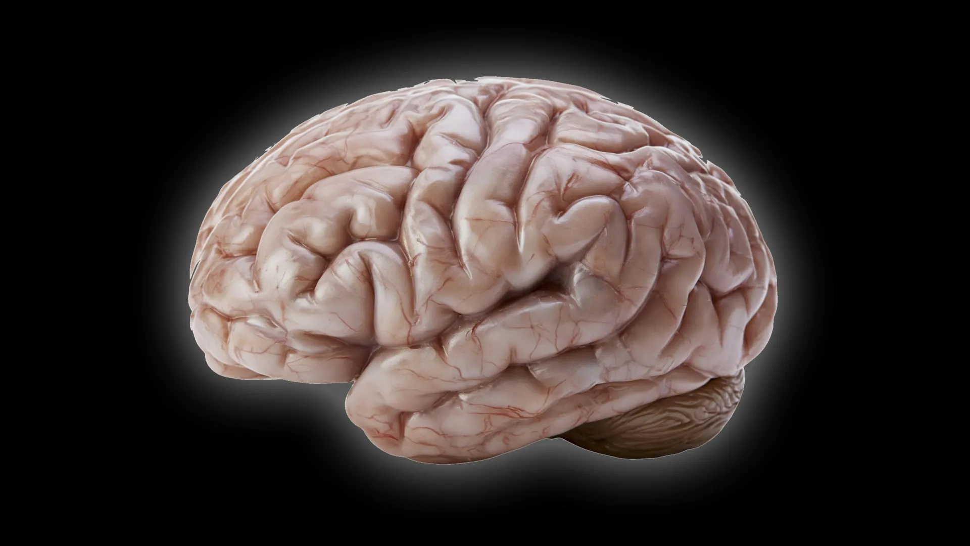
The diversity of brain morphology across the mammalian order of primates has long fascinated neuroscientists, evolutionary biologists, and anatomists alike. While the vast majority of living primates possess complex, enlarged cerebral cortices adorned with intricate systems of sulci (grooves) and gyri (folds), notable exceptions persist in nature. Chief among these anomalies is the common marmoset (Callithrix jacchus), a diminutive New World monkey native to the tropical forests of South America. Unlike humans, chimpanzees, or macaques—whose brains resemble tightly packed, folded walnuts designed to maximize surface area and cognitive capacity—the common marmoset features a brain that is almost entirely lissencephalic, or smooth.
For decades, the evolutionary mechanisms driving this dramatic divergence in brain structure remained largely obscure. However, recent empirical breakthroughs achieved by a team of researchers at the German Primate Center (DPZ)—the Leibniz Institute for Primate Research located in Göttingen, Germany—have begun to illuminate the microscopic processes responsible for these macro-level differences. By employing advanced stem cell technology, 3D organoid models, and comparative developmental biology, the DPZ research team has successfully identified specific cellular behaviors, structural variations, and chronological milestones that dictate why some primate brains undergo massive folding while others remain smooth. Published in the peer-reviewed journal Science Advances, the findings not only reshape our understanding of primate neuroanatomy and evolutionary trajectories but also establish a critical framework for investigating human neurodevelopmental disorders.
Main Facts and the Architectural Puzzle of Primate Cortices
To appreciate the significance of the German Primate Center’s findings, one must first examine the fundamental architecture of the mammalian brain. The cerebral cortex, the outer layer of neural tissue, is responsible for higher-order brain functions, including sensory processing, voluntary motor control, reasoning, and abstract thought. As evolutionary pressures drove the expansion of the primate brain over millions of years, surface area requirements quickly outpaced the physical constraints of the cranial vault. Nature solved this engineering challenge through cortical folding, a process known as gyrification. By creating folds and grooves, the brain packs an exponentially higher number of neurons into a constrained physical space, thereby enhancing computational power without necessitating a disproportionately massive skull.
Evolutionary anthropologists widely accept that the common ancestor of modern primates likely possessed a moderately sized brain exhibiting at least a foundational degree of folding. Yet, as lineages diverged, evolutionary adaptations took various paths. South American marmosets, which have become indispensable model organisms in modern biomedical and neuroscience research due to their small size, rapid reproductive cycle, and complex social behaviors, evolved in a direction that favored metabolic efficiency and miniaturization over extreme cortical expansion. Consequently, their brains retained a smooth, lissencephalic appearance.
The central mystery tackled by the DPZ research team was not merely that marmoset brains are smaller, but how the developmental program diverges from that of humans and other large-brained primates. "We wanted to understand which changes at the cellular level cause the marmoset brain to grow less and form fewer folds," explains Lidiia Tynianskaia, co-first author of the study and a doctoral candidate within the Junior Research Group Brain Development and Evolution at the DPZ. "At the beginning of development, the common marmoset brain exhibits the typical structure and composition of a large, folded primate brain. As development progresses, processes must therefore occur that effectively slow down the production of nerve cells."
Chronology and Methodology: The Journey from Cellular Models to Fetal Tissue
The investigation required a sophisticated methodological approach that bridged the gap between living tissue analysis and ethical constraints associated with primate research. Because studying early-stage neurogenesis directly in living primate embryos presents insurmountable ethical and logistical challenges, the research team turned to state-of-the-art stem cell technology.
By reprogramming somatic cells from humans and common marmosets into induced pluripotent stem cells (iPSCs), the scientists successfully cultivated cerebral organoids—often colloquially referred to as "mini-brains." These three-dimensional, lab-grown tissue structures replicate key architectural and biochemical properties of early human and primate embryonic brain development. Organoids provide an ideal experimental medium, enabling researchers to observe cellular dynamics across large sample sizes that would be statistically impossible to achieve using live animal subjects.
The chronology of the research unfolded across several distinct phases over the course of multiple years. Initially, the team focused on establishing reliable organoid cultures for both species. Once the cultures matured, the researchers performed meticulous comparative analyses using live-cell imaging, immunohistochemistry, and single-cell transcriptomics to monitor the behavior of neural progenitor cells—the foundational cells responsible for generating neurons.
A critical milestone in the project involved bridging the temporal gap between in vitro models and in vivo development. Organoids, while invaluable, do not always replicate the precise metabolic and temporal growth rates of natural embryonic tissue. To validate their findings, the research team meticulously mapped the developmental timeline of marmoset brain organoids against biological tissue samples. They discovered a precise temporal correlation: a marmoset brain organoid cultivated for 50 days corresponds structurally and developmentally to a natural marmoset embryonic brain at gestational day 90.
Armed with this validated calibration, the researchers confirmed their organoid-derived data by repeating key experiments using fetal brain tissue samples. This rigorous cross-verification ensured that the observed cellular phenomena were authentic reflections of developmental biology rather than artifacts of the in vitro culture environment.
Supporting Data: The Triad of Cellular Constraints
The empirical data gathered by the DPZ team revealed that the smooth, compact nature of the marmoset brain is not the consequence of a single genetic mutation or isolated structural alteration. Instead, it is the result of an intricate interplay of three distinct cellular limitations operating in tandem.
First, the researchers discovered marked differences in cell division rates. Certain neural progenitor populations within the marmoset model divide at a significantly slower pace than their human counterparts. "Our investigations have shown that certain progenitor cells in the common marmoset divide significantly more slowly than in humans," notes César Mateo Bastidas Betancourt, the study’s co-first author and a PhD student within the same DPZ research group.
Second, structural disparities among progenitor cells heavily influence proliferative capacity. Progenitor cells in marmosets possess a simpler morphological structure, featuring noticeably fewer cellular processes (extensions or projections) than equivalent human progenitor cells. This structural simplicity correlates directly with reduced proliferative output, meaning each progenitor gives rise to fewer daughter cells over its lifecycle.
Third, the temporal window of rapid cell multiplication is substantially compressed in marmosets compared to humans. The developmental window during which progenitor cells undergo rapid, expansive symmetric and asymmetric divisions is significantly shorter in the South American primates.
Together, this triad of constraints—slower cell division, simpler cellular architecture, and a contracted developmental timeline—acts as a biological brake on neurogenesis. By throttling the total volume of neurons produced during embryonic development, the marmoset brain avoids the massive cellular accumulation that physically forces cortical folding in larger-brained mammals.
Official Responses and Academic Implications
The publication of these findings in Science Advances has drawn positive reactions from the broader neurodevelopmental and evolutionary biology communities. Independent experts have praised the study for successfully demonstrating how subtle micro-level tweaks in cellular kinetics can accumulate to produce profound macro-evolutionary divergence.
Dr. Michael Heide, head of the Junior Research Group Brain Development and Evolution at the DPZ and senior author of the study, emphasized the methodological triumph of combining laboratory models with biological validation. "The study combines the advantages of in vivo and in vitro methods," Heide stated. "Organoids are well-suited for obtaining statistically robust results because such sample sizes are not feasible in primates. We subsequently repeated some key experiments in fetal brain tissue to confirm the results from the organoids."
From an academic perspective, the research provides concrete empirical data to support long-standing theoretical models regarding brain evolution. Historically, scientists debated whether lissencephaly in smaller mammals was an ancestral trait retained from primitive mammalian precursors or a secondary evolutionary adaptation favoring compact size. By demonstrating that the marmoset brain initiates development with structural markers typical of folded brains before actively deploying cellular brakes, the DPZ study strongly supports the hypothesis that smooth brains can evolve as specialized secondary adaptations.
Broader Impact and Clinical Relevance for Human Health
While the immediate value of the research lies in evolutionary biology and comparative neuroanatomy, the broader implications extend deeply into clinical medicine and human health. The fundamental cellular mechanisms governing progenitor cell proliferation, cell cycle duration, and morphological complexity are largely conserved across primates, including Homo sapiens.
Understanding the precise molecular and cellular levers that control neuron production provides an essential baseline for human neurodevelopmental research. Human neurodevelopmental disorders—such as microcephaly (abnormally small brains and skulls), lissencephaly syndromes, and certain forms of autism and intellectual disability—frequently involve disruptions in neural progenitor proliferation, cell division timing, or structural migration during embryogenesis.
By isolating the exact cellular processes that naturally throttle brain growth and folding in marmosets, scientists gain a sharper contrastive tool for identifying what goes awry when human brain development is disrupted by genetic mutations or environmental teratogens. If researchers can pinpoint how evolution successfully dialed down neuron production in the marmoset lineage, they may better understand the pathological mechanisms that prematurely halt cortical expansion in human disease states.
Furthermore, as the common marmoset continues to rise in prominence as a non-human primate model for translational neuroscience—valued for its genetic tractability and complex behavioral repertoire—a comprehensive understanding of its basic neurodevelopmental biology is indispensable. Researchers utilizing marmosets in laboratories worldwide must account for these fundamental structural and developmental divergences when interpreting neurological data and translating animal models to human clinical applications.
Conclusion: A New Chapter in Primate Neurobiology
The investigation conducted by the German Primate Center successfully demystifies one of nature’s elegant anatomical variations. By drilling down from macroscopic brain morphology to microscopic organoid cultures and fetal tissue dynamics, the research team mapped the precise cellular choreography that keeps the common marmoset’s brain smooth and compact. Through the interplay of slower cell division rates, simpler progenitor architecture, and shortened developmental windows, nature achieves a distinct neuroanatomical blueprint tailored to the evolutionary niche of a small arboreal primate.
As science continues to probe the vast landscape of mammalian evolution, studies of this caliber underscore the profound power of comparative developmental biology. By examining the exceptions to the rule—such as the smooth-brained marmoset—researchers not only shed light on the diverse evolutionary pathways carved across millions of years, but also secure critical insights into the fragile, intricate mechanisms that construct the human mind.
