
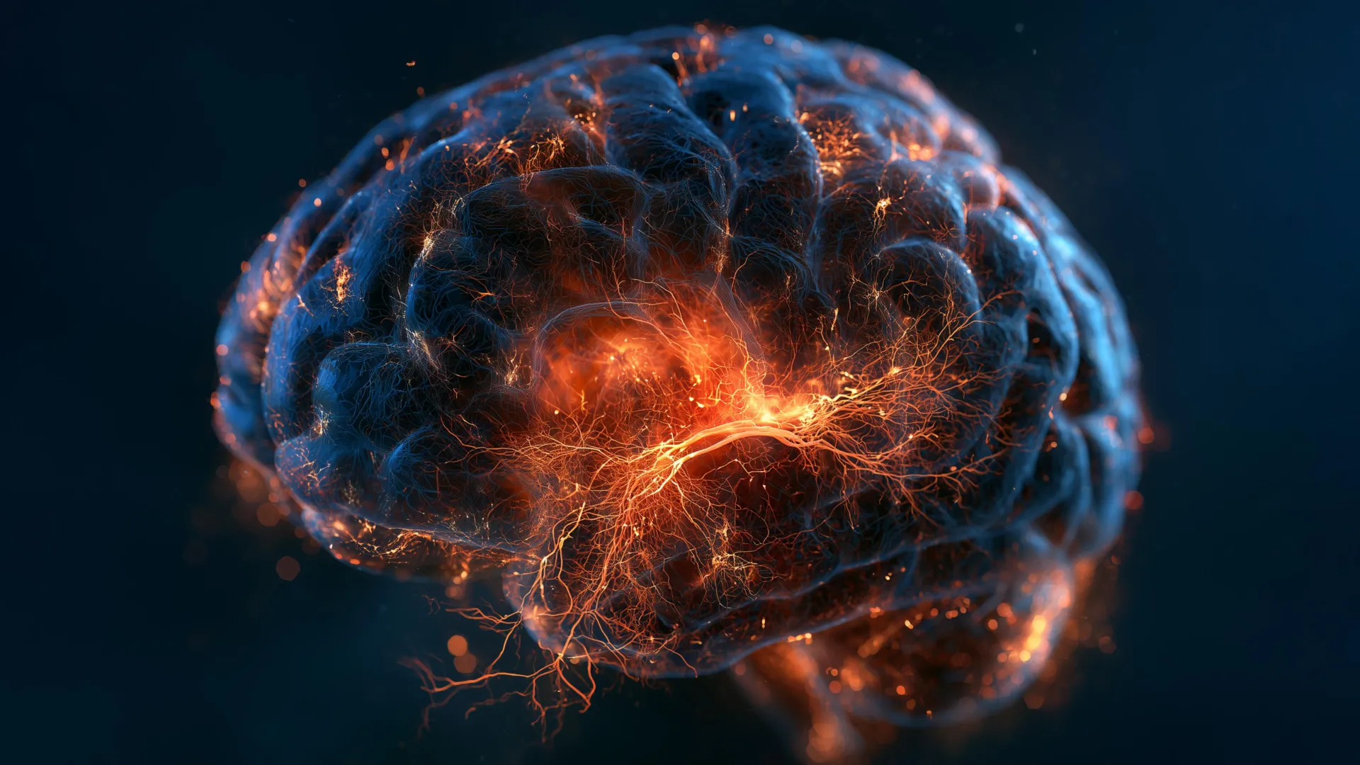
The human brain is traditionally understood as a static landscape after early childhood development, possessing a fixed complement of roughly 100 billion neurons established primarily before birth. However, groundbreaking new research led by scientists at the Columbia University Vagelos College of Physicians and Surgeons has demonstrated that a select region of the adult brain continues to generate fresh neural cells—and that this vital biological process stalls entirely in individuals suffering from major depressive disorder (MDD). Published on August 21, 2026, in the peer-reviewed journal Nature Medicine, the study titled "Dysregulated adult hippocampal neurogenesis in major depressive disorders" offers the first direct empirical evidence of halted neurogenesis in human adults with clinical depression. By mapping nearly half a million individual brain cells, the research team not only illuminated a core physical mechanism underlying mood disorders but also established a molecular blueprint that could fundamentally transform how psychiatry diagnoses and treats mental illness.
Main Facts and Core Findings
For decades, psychiatric medicine relied heavily on the monoamine hypothesis of depression, which conceptualized the disorder primarily as a chemical imbalance involving neurotransmitters such as serotonin, norepinephrine, and dopamine. While medications targeting these neurotransmitter systems—such as selective serotonin reuptake inhibitors (SSRIs)—have helped millions of patients, they frequently fail to provide adequate relief for treatment-resistant cases, and their precise mechanisms of action have remained incompletely understood.
The Columbia University study shifts the paradigm toward neuroplasticity, suggesting that depression is fundamentally a disease of cellular adaptation. Led by Dr. Maura B. Dupont, professor of psychiatry at Columbia and senior author of the study, the research team discovered that the generation of new neurons—known as adult neurogenesis—ceases or severely stalls in the hippocampus of individuals with major depressive disorder.
The hippocampus, a seahorse-shaped structure nestled deep within the brain’s temporal lobes, is critical for consolidating episodic memories, regulating emotional responses, and contextualizing environmental stimuli. It is also one of the rare anatomical niches in the adult mammalian brain that retains the capacity to produce new neurons throughout life. According to the research findings, the inability to generate these newborn cells deprives patients of the cognitive flexibility and resilience necessary to adapt to chronic stress and changing life circumstances.
To arrive at these conclusions, Dr. Dupont’s laboratory conducted an exhaustive single-cell and molecular analysis of nearly 500,000 individual brain cells procured post-mortem from donors diagnosed with major depressive disorder, alongside healthy control subjects. Utilizing advanced transcriptomic and proteomic methodologies, the team analyzed gene expression patterns, cellular signaling pathways, and protein alterations within specific micro-circuits of the hippocampus.
The data revealed widespread molecular disruptions extending far beyond simple neuron counts. The trisynaptic circuit—the primary neural pathway through which the hippocampus processes and encodes emotional memories—exhibited marked signs of neuroinflammation, metabolic stress, and cellular dysfunction. Genes responsible for forging new synaptic connections, facilitating intercellular communication, supplying cellular energy, and transporting intracellular materials were all found to be dysregulated in depressed brains.
Chronology and Research Methodology
The scope of this landmark study represents the culmination of years of technological advancement in single-cell genomics, proteomics, and computational biology. The scientific timeline leading to this publication reflects a gradual convergence of animal model research, post-mortem human tissue analysis, and high-throughput data processing.
For decades, neuroscientists debated whether adult neurogenesis occurred in humans to the same degree observed in rodents. While initial rodent studies confirmed that new neurons were born in the subgranular zone of the hippocampal dentate gyrus and played a direct role in cognitive functions like pattern separation, validating these findings in human tissue remained technically intractable. Traditional bulk tissue analysis homogenized thousands of distinct cells, masking subtle abnormalities occurring within specialized cellular micro-environments.
The breakthrough for the Columbia team came through the integration of multiple state-of-the-art platforms housed at the medical center:
- Single-Cell RNA Sequencing: Performed at the JP Sulzberger Columbia Genome Center, allowing researchers to measure the activity of every single gene within individual cells.
- Proteomic Profiling: Conducted at the Columbia University Department of Biology’s Quantitative Proteomics and Metabolomics Center to examine whether cellular proteins had undergone pathological alterations.
- Computational Clustering: Executed via Columbia’s Center for Computational Biology and Bioinformatics to map the precise anatomical and functional locations of affected cells within the hippocampal circuit.
By analyzing post-mortem brain samples from carefully screened cohorts, the research team systematically mapped the cellular architecture of depression. This meticulous workflow bridged the gap between behavioral psychology and microscopic molecular biology, providing a high-resolution window into the human brain previously unavailable to psychiatric researchers.
Supporting Data and the Mechanics of Memory
To understand the profound clinical implications of halted neurogenesis, researchers focused closely on a cognitive process known as pattern separation. Pattern separation is the brain’s ability to distinguish between similar yet distinct memories and to separate the emotional weight of past events from current experiences.
In healthy individuals, newborn neurons in the hippocampus integrate rapidly into existing memory circuits. Because these immature neurons are exceptionally plastic and responsive to novel stimuli, they facilitate the accurate archiving of new memories, preventing them from bleeding into older, emotionally charged recollections.
When neurogenesis stalls, pattern separation breaks down. Individual memories and their associated emotional contexts blur together. Dr. Dupont illustrated this phenomenon through a common clinical observation: an individual interacting with a tired, quiet friend might, under normal circumstances, recognize the interaction as a unique, neutral event. However, an individual with impaired pattern separation fails to separate the current context from historical memories of rejection or trauma, erroneously concluding, "They are upset with me" or "I am being rejected."
"You may be out with a friend for lunch, but she’s tired and doesn’t talk much," Dupont explained. "With intact pattern separation, you remember this as a unique event. With impaired pattern separation, it becomes mixed with previous memories of feeling rejected… And I see this a lot in my patients, where they can only retrieve negative information from their memories."
This cognitive bias—the tendency to preferentially retrieve negative autobiographical information while failing to encode neutral or positive context—fuels the ruminative cycles characteristic of clinical depression. Animal studies and clinical observations from patients who underwent targeted radiation therapy for brain tumors (which inadvertently halted hippocampal neurogenesis) have consistently supported the hypothesis that newborn neurons are functionally indispensable for maintaining this cognitive boundary.
Epigenetics, Environmental Factors, and Genetic Diversity
Beyond cellular counts and neurogenesis rates, the Columbia study investigated the upstream genetic and epigenetic drivers of these hippocampal abnormalities. The research team identified altered expression in several specific genes whose genetic variants have long been linked to major depressive disorder in genome-wide association studies (GWAS).
Crucially, the study also uncovered widespread epigenetic modifications among the disrupted genes. Epigenetic mechanisms do not alter the underlying DNA sequence itself; instead, they act as molecular dimmer switches—such as DNA methylation and histone modification—that dictate how strongly specific genes are turned on or off. These epigenetic tags are highly sensitive to environmental pressures, including chronic psychological stress, early-life trauma, learning, aging, and exposure to environmental chemicals.
"These are like dimmer switches that control how active genes are, and they are affected by life experiences such as stress, learning, aging, chemicals, etc.," noted Dupont.
This complex interplay between innate genetic vulnerability and environmental adversity helps explain why major depressive disorder manifests with such extraordinary clinical heterogeneity. Two patients diagnosed with clinical depression may display entirely different symptom profiles—ranging from insomnia, anhedonia, and psychomotor retardation to severe cognitive fatigue and anxiety—because the underlying molecular disruptions within their hippocampal circuits are fundamentally distinct.
"Overall, the wide range of effects we found could reflect different pathogenetic mechanisms, perhaps indicating that depression is not just one disease," Dupont added.
Broader Impact and Implications for Precision Psychiatry
The publication of this study arrives at a pivotal juncture in the evolution of mental healthcare. For generations, psychiatry has operated under a diagnostic framework based primarily on behavioral symptom checklists outlined in the Diagnostic and Statistical Manual of Mental Disorders (DSM). While this approach provides clinical utility, it groups biologically distinct conditions under single diagnostic umbrellas, contributing to high trial-and-error rates in pharmacological treatment.
The Columbia University findings lay the scientific groundwork for a radical transition toward precision psychiatry, mirroring the trajectory of modern oncology. Over the past several decades, cancer treatment shifted away from categorizing tumors merely by their anatomical location (e.g., lung cancer or breast cancer) toward classifying them by their underlying molecular and genetic signatures (e.g., EGFR-mutant or HER2-positive). This molecular taxonomy enabled the development of targeted therapies that dramatically improved patient outcomes.
Dr. Dupont and her colleagues envision a comparable revolution for psychiatric medicine. By identifying distinct molecular subtypes of depression—such as those driven primarily by impaired adult neurogenesis versus those characterized by localized neuroinflammation or synaptic connectivity deficits—clinicians could eventually tailor therapeutic interventions to the specific biological failure occurring within a patient’s brain.
"We want to reclassify depression based on its molecular features, similar to what has been done in cancer," Dupont stated. "Classifying cancers based on their cellular characteristics, not their locations, has led to new and improved treatments. We hope the same will be true for depression and other psychiatric or brain diseases."
Therapeutically, the discovery offers a compelling roadmap for drug development. If pharmacological agents or neuromodulatory techniques can be engineered to safely reactivate adult neurogenesis and restore structural plasticity within the human hippocampus, clinicians may soon possess the tools not merely to manage the symptoms of depression, but to structurally rewire the neural circuits damaged by chronic stress.
Study Authorship and Institutional Support
The expansive research effort was conducted collaboratively within the Maura Dupont Laboratory at Columbia University Irving Medical Center and the New York State Psychiatric Institute.
The comprehensive author list accompanying the Nature Medicine publication includes Madeleine S. Peng, Jialin Jiang, Lucia Polizzi, Tiancheng Shi, Rakshitha Ramkumar, Victor O. Anosike, Giulia Guasoni, Alexandra M. Wamalwa, Madeline B. Mariani, Cheick A. Sissoko, Alexandria N. Tartt, Camille Fulmore, Gorazd B. Rosoklija, Yung-yu Huang, Victoria Arango, Shujuan T. McDonald, Natasha Bitoljanu (representing Ss. Cyril and Methodius University in Macedonia), Joseph J. Mann, Phi T. Nguyen, Andrew J. Dwork, Lewis M. Brown, René Hen, Hanga Galfalvy, and Maura B. Dupont. Most contributing researchers maintain dual affiliations with Columbia University and the New York State Psychiatric Institute.
Institutional infrastructure played a vital role in executing the multi-layered analyses required for the project. In addition to sequencing conducted at the JP Sulzberger Columbia Genome Center and computational clustering performed through Columbia’s Center for Computational Biology and Bioinformatics, advanced proteomic evaluations were delivered by the Quantitative Proteomics and Metabolomics Center within the Columbia University Department of Biology.
As the medical community digests these findings, future research will focus on translating these post-mortem cellular insights into living biomarkers and targeted clinical interventions. While substantial work remains to fully map the human neurogenic mechanism from end to end, Columbia University’s landmark study marks a definitive turning point in our scientific comprehension of depression—moving humanity closer to a future where mental illness is understood, diagnosed, and cured at the level of the individual cell.
