
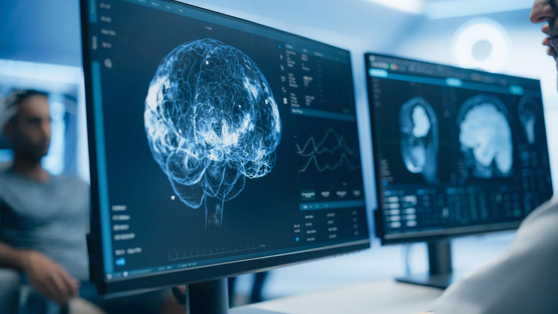
A groundbreaking brain imaging study originating from the University of Turku in Finland is poised to reshape our understanding of long COVID, a condition that continues to affect millions globally. For an extended period, a leading hypothesis for the persistent, debilitating symptoms associated with long COVID has centered on ongoing inflammation within the brain, directly linked to the SARS-CoV-2 virus. However, this new research presents compelling evidence that challenges this prevailing narrative, indicating a lack of widespread brain inflammation in individuals experiencing lingering post-infection symptoms. Instead, the study’s findings point towards altered activity in brain regions crucial for emotion, stress response, and memory as a more likely contributor to the severity of symptoms.
The implications of these findings are significant, potentially redirecting research efforts and the development of therapeutic strategies for a condition that has defied easy explanation and treatment since its emergence in late 2019 and early 2020. As the scientific community grappled with the initial COVID-19 pandemic, the long-term sequelae, now commonly referred to as long COVID or post-acute sequelae of SARS-CoV-2 infection (PASC), began to emerge as a distinct and formidable public health challenge. Symptoms such as profound fatigue, cognitive dysfunction often described as "brain fog," persistent anxiety, and depression have become hallmarks of this condition, prompting extensive investigation into their underlying biological mechanisms.
Challenging the Inflammation Paradigm
The theory of ongoing neuroinflammation as the primary driver of long COVID symptoms gained traction due to observations of inflammatory markers in some individuals during the acute phase of infection and in certain animal models. Early neuropathological studies of severe acute COVID-19 cases did indeed reveal clear signs of inflammation in brain tissue. This led many researchers to hypothesize that residual viral activity or the body’s sustained immune response could lead to chronic inflammatory processes in the central nervous system, thereby disrupting normal brain function and manifesting as the diverse array of long COVID symptoms. However, direct, robust evidence specifically demonstrating widespread, persistent neuroinflammation in the majority of long COVID patients has remained elusive.
To address this critical gap in knowledge, a team of researchers at the University of Turku, renowned for its expertise in neuroimmunology, embarked on a comprehensive investigation. Led by Professor of Neuroimmunology and InFLAMES Research Flagship group leader Laura Airas, the study employed state-of-the-art brain imaging techniques to meticulously examine the brains of individuals who continued to suffer from long COVID symptoms well after their initial SARS-CoV-2 infection had subsided. The InFLAMES (Immune System and Neuroinflammation) Research Flagship is a collaborative initiative between the University of Turku and Ã…bo Akademi University, dedicated to advancing the understanding of immune system interactions with the nervous system and developing novel diagnostic and therapeutic approaches.
"We did not observe evidence of widespread brain inflammation in patients with long COVID when compared to healthy controls," stated Professor Airas, underscoring a key finding that directly contradicts the prevailing inflammation-centric hypothesis. This assertion is based on rigorous scientific methodology and comparative analysis.
A Comparative Study Design
The study’s robust design involved a comparative approach, including three distinct participant groups to provide crucial context and benchmarks. The cohort comprised 14 individuals diagnosed with long COVID who were experiencing persistent symptoms, 11 healthy volunteers serving as a control group, and 13 individuals diagnosed with multiple sclerosis (MS). MS was included as a comparator group because it is a well-established neurological disease characterized by significant and well-documented inflammation within the brain’s white matter. This inclusion allowed researchers to gauge the level of neuroinflammation in long COVID patients relative to a known inflammatory neurological condition.
All participants underwent a series of advanced neuroimaging and biological analyses. Positron Emission Tomography (PET) scans were utilized, specifically designed to detect and quantify neuroinflammation by identifying areas of heightened metabolic activity associated with immune cell activation. Complementing the PET scans were Magnetic Resonance Imaging (MRI) scans, which provided detailed structural information about the brain and allowed for the assessment of changes in white matter integrity – a common area affected by inflammation in neurological disorders. Furthermore, blood samples were collected and analyzed for specific biological markers that indicate neuronal damage and the health of supporting brain cells, known as glial cells.
Key Findings: Inflammation Levels and Temporal Dynamics
The results of these comprehensive analyses yielded several significant insights. When comparing the long COVID group with the MS patients, researchers found markedly lower levels of inflammatory activity in the brain’s white matter among those with long COVID. This stark difference suggests that the type and extent of neuroinflammation observed in MS, a condition with a clear inflammatory pathology, are not replicated in the long COVID cohort investigated.
Crucially, when long COVID patients were compared to the healthy volunteers, researchers detected no meaningful differences in biological markers associated with brain inflammation or neurodegeneration. This absence of elevated inflammatory markers or signs of neuronal breakdown in the blood samples further supports the PET scan findings and casts doubt on the widespread inflammatory hypothesis for long COVID.
However, the study did uncover a nuanced temporal aspect to inflammation. Previous neuropathological studies focusing on the acute phase of severe COVID-19 have reported clear inflammatory signatures in brain tissue. In this new study, a pattern emerged regarding the timing of infection. Participants who were scanned within 16 months of their initial COVID-19 infection exhibited slightly higher levels of inflammatory activity in their white matter compared to those who had been sick for a longer duration.
Professor Airas suggests that this observation may indicate that inflammation, if present, is more pronounced in the earlier stages following SARS-CoV-2 infection and gradually diminishes over time. This temporal dynamic implies that while some level of inflammation might occur acutely, it does not necessarily persist as a widespread, driving force for long-term symptoms. This aligns with the understanding that many viral infections can trigger transient inflammatory responses that resolve as the body recovers.
Shifting Focus to Emotion and Stress Pathways
Beyond the inflammation findings, the study revealed another critical pattern that offers a potential alternative explanation for persistent long COVID symptoms. Researchers observed a significant correlation between symptom severity and increased cellular activity in specific brain regions. Patients who reported higher levels of anxiety and depression, and who consequently experienced a poorer quality of life, displayed heightened activity in the hippocampus and the amygdala.
The hippocampus is a brain structure critically involved in learning, memory formation, and spatial navigation. The amygdala, on the other hand, is a key component of the limbic system, playing a central role in processing emotions, particularly fear and anxiety, and in mediating the body’s stress response. The increased activity in these regions among symptomatic long COVID patients suggests a potential dysregulation of emotional processing and stress reactivity, which could manifest as many of the subjective complaints reported by individuals with long COVID.
The researchers posit that these findings strongly suggest that altered activity within these emotion-related and stress-response brain areas could be intrinsically linked to the severity of the symptoms experienced by some individuals with long COVID. This perspective shifts the focus from a generalized inflammatory insult to a more nuanced understanding of how the brain’s emotional and stress-related circuits might be affected.
Implications for Future Treatments
The implications of this research are far-reaching and could significantly influence the trajectory of future long COVID treatment strategies. The findings from Professor Airas and her team provide a more refined scientific understanding of long COVID and directly challenge the long-held notion that persistent brain inflammation is the primary culprit behind prolonged symptoms in all affected patients.
Instead, this study advocates for a more complex model of the condition, one in which inflammatory changes may be most prominent in the immediate aftermath of infection and subsequently wane. This recalibration of understanding is crucial for developing effective interventions.
Long COVID remains a formidable global health challenge, impacting the lives of millions worldwide. The chronic and often debilitating nature of its symptoms can persist for months, and in some cases, even years, after the initial viral illness. This new research offers a hopeful direction for those seeking relief. Based on these findings, it is plausible that a subset of patients with persistent symptoms might find greater benefit from therapeutic approaches that target stress management and emotional regulation rather than solely relying on anti-inflammatory therapies.
"This study highlights the need to continue investigating the complex biological mechanisms underlying long COVID," Professor Airas emphasized. "Understanding these processes is essential for developing targeted treatments." This underscores the ongoing commitment to unraveling the intricate biology of this post-viral syndrome.
The research, conducted by Professor Airas and her colleagues, was published in the peer-reviewed Journal of Neurology, a respected publication in the field, lending credibility and visibility to its findings. The InFLAMES Flagship, a cornerstone of this research, represents a significant investment by Finland in interdisciplinary scientific exploration, aiming to bridge the gap between immunology and neurological research to pioneer new diagnostic tools and personalized medical treatments. This initiative is supported by the Research Council of Finland’s Flagship Program, underscoring the national commitment to addressing critical health challenges.
Broader Impact and Next Steps
The study’s findings have the potential to catalyze a paradigm shift in how long COVID is understood and treated. By moving beyond a singular focus on inflammation, researchers can now explore a broader spectrum of neurobiological dysfunctions, including those related to neuroendocrine pathways, autonomic nervous system regulation, and indeed, the intricate interplay of emotion and cognition. This more comprehensive view may lead to the development of personalized treatment plans, recognizing that long COVID is likely a heterogeneous condition with diverse underlying mechanisms contributing to its varied symptomatology.
For patients, this research offers a glimmer of hope. It suggests that current therapeutic avenues may not be fully optimized and that alternative strategies, focused on restoring balance in the brain’s emotional and stress response systems, could be more effective for certain individuals. This could involve exploring interventions such as cognitive behavioral therapy (CBT) tailored for long COVID symptoms, mindfulness-based stress reduction techniques, and potentially pharmacological agents that modulate neurotransmitter systems involved in mood and anxiety.
The scientific community will undoubtedly build upon these findings. Future research will likely aim to replicate these results in larger, more diverse cohorts, explore the specific mechanisms by which emotional and stress pathways become dysregulated, and investigate the long-term effects of interventions targeting these areas. Understanding the temporal evolution of these changes and identifying biomarkers that can predict which patients are more likely to benefit from specific treatments will be critical next steps.
In conclusion, this pioneering study from the University of Turku represents a significant step forward in the ongoing battle against long COVID. By challenging established assumptions and illuminating new avenues of investigation, it paves the way for a more nuanced, effective, and ultimately, more hopeful approach to managing this complex and pervasive post-viral syndrome. The focus is shifting from a singular fight against inflammation to a more holistic understanding of the brain’s intricate systems, offering a renewed promise for recovery for millions affected worldwide.
