
For decades, the mainstream exploration of neurobiology has operated under a genetic and protein-centric paradigm. Laboratories worldwide have dedicated monumental resources to sequencing genomes, charting neural circuitry, cataloging cell types, and tracking the intricate signaling proteins that govern how neurons fire and communicate. Yet, nestled within the complex machinery of the central nervous system lies a profoundly influential component that has historically defied systematic cartography: lipids. These essential fatty molecules form the foundational fluid mosaic of the membranes enveloping every neuron, construct the protective myelin sheaths that insulate long-range nerve fibers, and act as dynamic mediators in intercellular signaling cascades. Despite their ubiquitous presence and biological indispensability, lipids have remained a notoriously blurry variable in the neurological equation.
This persistent blind spot in neuroscience has recently begun to clear. A pioneering team of researchers at the École Polytechnique Fédérale de Lausanne (EPFL) has officially bridged this historical divide, constructing the first comprehensive, high-resolution three-dimensional map of lipid distribution across the mouse brain. Published in the prestigious journal Nature, this landmark achievement introduces the "Lipid Brain Atlas"—an open-access digital resource that reveals an astonishing degree of chemical organization previously invisible to conventional anatomical and genetic methodologies. Spearheaded by Professor Giovanni D’Angelo, alongside core contributors Luca Fusar Bassini and Gioele La Manno, the project fundamentally alters our understanding of brain architecture, exposing a hidden layer of chemical geography that shifts dramatically during critical life events such as pregnancy and holds profound implications for neurodegenerative pathologies.
The Core Breakthrough: Unveiling 539 Chemical Territories
To fully appreciate the magnitude of the EPFL breakthrough, one must examine the technical hurdles that have stymied lipid mapping for generations. Unlike proteins and nucleic acids, which can be easily tagged, amplified, or sequenced using standard molecular biology toolkits, lipids are chemically diverse, structurally similar, and notoriously labile. Conventional imaging techniques often struggle to distinguish individual lipid molecules or preserve their precise spatial orientation within delicate tissue samples. Consequently, neuroscientists have long possessed detailed atlases of gene expression and cellular morphology, while remaining functionally blind to the chemical lipid topography that underpins those very structures.
To overcome these barriers, the EPFL research collective deployed advanced mass spectrometry imaging. This sophisticated analytical technique allows scientists to precisely interrogate thin slices of biological tissue using a focused laser beam. As the laser systematically scans across each slice location by location, it vaporizes and ionizes the microscopic constituents, generating a detailed mass spectrum that reveals precisely which lipid species are present and in what quantities.
The scope of the undertaking was vast. The research team meticulously analyzed 172 distinct lipid molecules across 109 individual brain slices harvested from 11 test subjects. This rigorous processing pipeline yielded approximately 7 million distinct spatial and chemical measurements. To transform this massive numerical dataset into a coherent, navigable format, the investigators integrated custom machine learning algorithms designed specifically to handle the multi-dimensional complexity of lipid spatial distribution.
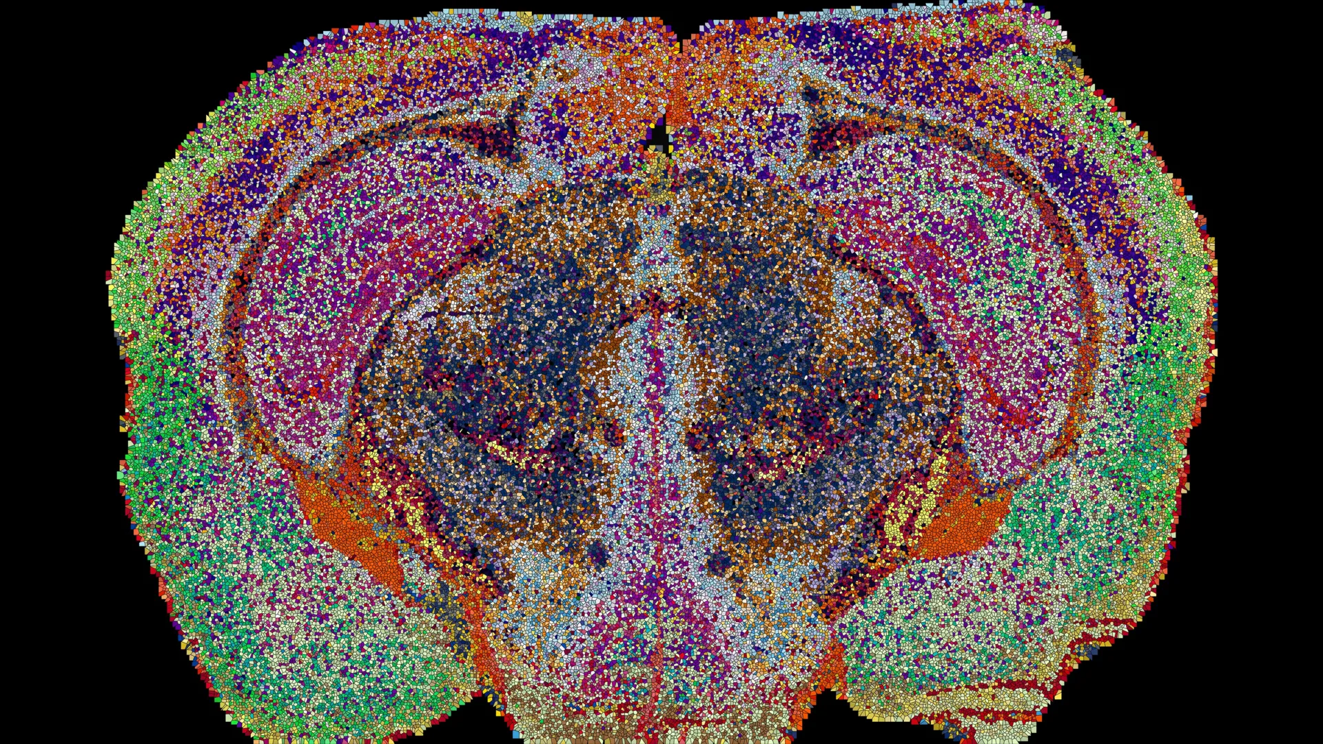
The resulting 3D reconstruction exposed a breathtakingly intricate landscape. The EPFL team identified 539 chemically distinct territories within the mouse brain, a classification they have termed "lipizones." Each lipizone possesses a unique chemical signature characterized by a specific ratio and composition of lipid molecules. Remarkably, these boundaries are so distinct and reproducible that researchers could accurately determine the anatomical origin of an isolated brain tissue sample simply by analyzing its lipid profile. While many of these newly mapped territories aligned neatly with classical neuroanatomical structures and recognized cell populations, a significant portion defied traditional categorization.
Methodology and Chronology of the EPFL Project
The creation of the Lipid Brain Atlas was the culmination of years of interdisciplinary collaboration, combining advanced analytical chemistry, high-throughput spatial biology, and machine learning computation. While the exact timeline of the project spans several years of optimization, instrument calibration, and data validation, the collaborative synergy between EPFL’s diverse departments proved decisive.
In the initial phase, the team focused on standardizing the tissue collection and mass spectrometry protocols to minimize degradation and maximize spatial fidelity. Brain tissue preservation is notoriously difficult when studying lipids, as these molecules can easily diffuse or degrade post-mortem. By refining rapid-freezing techniques and optimizing the matrix application required for matrix-assisted laser desorption/ionization (MALDI) mass spectrometry, the researchers ensured that the spatial localization of delicate membrane lipids remained intact.
Once the physical acquisition pipeline was established, the team processed the iterative slices to compile raw spectral data. The sheer volume of information—scaling into the millions of data points per brain—necessitated the development of specialized computational pipelines. Standard neuroimaging software is typically built to handle voxel-based structural MRI or genetic expression coordinates; it is fundamentally unequipped for the high-dimensional chemical matrices generated by lipidomics. By engineering bespoke machine learning clustering algorithms, the EPFL bioinformaticians successfully segmented the 3D reconstructions into the 539 distinct lipizones, cross-referencing their findings against established Allen Brain Atlas datasets to validate anatomical correlations.
Redrawing the Map: Surprising Revelations in White Matter and Beyond
One of the most provocative revelations of the Lipid Brain Atlas is its challenge to long-held assumptions regarding tissue uniformity. Neuroscientists have traditionally viewed certain regions of the central nervous system, particularly white matter—the dense network of myelinated nerve fibers that acts as the brain’s information highway—as relatively homogeneous in its chemical composition. After all, white matter’s primary role is insulation and signal transmission, duties traditionally attributed to a uniform coating of myelin.
The lipid atlas shattered this perception. The EPFL team discovered that white matter is actually compartmentalized into numerous distinct lipid zones. This surprising heterogeneity indicates that the oligodendrocytes and other glial cells responsible for synthesizing and maintaining myelin are far more chemically specialized than their genetic profiles alone would suggest.
Furthermore, the mapping initiative uncovered previously unrecognized chemical boundaries within the choroid plexus—the specialized network of cells responsible for producing cerebrospinal fluid—as well as along the ependymal tissues lining the brain’s internal ventricular cavities. These discoveries demonstrate that lipid mapping provides an orthogonal layer of biological information that complements, and frequently expands upon, data derived solely from genomics and transcriptomics.
In describing these functional relationships, Professor D’Angelo offers a compelling analogy: "In effect, lipids work like postal codes, giving distant but related parts of the brain the same chemical address." This chemical addressing system frequently bridges anatomically disparate regions that share functional circuits, revealing structural and chemical relationships that remain entirely invisible when researchers rely exclusively on gene activity maps.
Pregnancy and Plasticity: Widespread Shifts in Neural Chemistry
Beyond mapping the baseline architecture of the healthy adult brain, the research team sought to investigate how this chemical landscape responds to physiological stress and systemic biological transitions. To this end, the investigators applied their mass spectrometry imaging pipeline to examine the brains of pregnant mice, capturing a snapshot of neurochemical adaptation during gestation.
The results were striking. The analysis revealed profound, widespread shifts in lipid composition across numerous brain regions during pregnancy, outstripping the baseline lipid variations typically observed between male and female subjects. The cerebral cortex, the outermost layer of the brain responsible for higher-order cognitive processing, sensory perception, and motor control, exhibited particularly extensive chemical reorganization.
Concurrently, researchers observed a sharp, systemic surge in the levels of galactosyl ceramide—a critical sphingolipid and essential structural component of myelin—throughout much of the brain’s white matter. These sweeping alterations suggest that the maternal brain undergoes a coordinated chemical retuning to accommodate the physiological, metabolic, and behavioral demands of pregnancy.
"Together, these shifts suggest the brain retunes its own wiring to meet the demands of pregnancy," noted co-investigator Gioele La Manno. "The Atlas reveals that the brain constantly adjusts its lipids to meet the body’s changing needs." This discovery opens an entirely new avenue of investigation into maternal neuroplasticity, suggesting that structural adaptations during gestation involve fundamental membrane remodeling rather than merely functional synaptic changes.
Broader Implications for Health, Disease, and Aging
The public release of the Lipid Brain Atlas as a freely accessible online resource represents a major milestone for the global neuroscience community. By allowing researchers worldwide to interrogate this digital repository, EPFL has effectively democratized lipidomic research, providing a robust reference baseline for healthy mouse brain tissue.
The implications of this baseline extend far beyond basic neuroanatomy. Historically, researchers have noted significant alterations in lipid metabolism and membrane composition in various neurological and psychiatric conditions, ranging from major depressive disorder to Alzheimer’s disease. However, without a high-resolution map of normal lipid distribution, distinguishing pathological aberrations from physiological variations has proved immensely challenging. With the new atlas, scientists now possess a high-resolution reference tool to investigate how disruptions in lipid localization contribute to neurodevelopmental disorders, cognitive decline during aging, and neurodegenerative pathology.
While the current map is derived from murine models and cannot be directly translated to human clinical diagnostics without extensive further study, the underlying methodological framework is universally applicable. As Luca Fusar Bassini emphasized, "What transfers to humans isn’t the map, it’s the method, and the fact that we now know that a healthy brain has a very organized lipid map at all."
As the scientific community begins to digest these findings, the EPFL lipid atlas marks the dawn of a new era in neurobiology—one where lipids are no longer treated as passive structural filler, but as dynamic, highly organized codes that dictate the very structure, adaptation, and vulnerability of the mind.
