
The pursuit of mastery has long baffled educators, clinicians, and cognitive scientists alike. Why do certain physical and cognitive skills seem to anchor themselves into muscle memory almost instantly, while others remain stubbornly out of reach despite hours of painstaking repetition? Traditional pedagogical and psychological frameworks have largely attributed these discrepancies to innate talent, cognitive aptitude, or the sheer volume of deliberate practice. However, a growing body of neuroscientific research suggests that these factors, while undeniably important, tell only part of the story. The missing variable may lie not within the mind alone, but in the physiological state of the entire organism—specifically, whether the brain is biologically primed to convert transient practice into permanent neural architecture.
In a significant breakthrough published in the journal iScience on August 25, 2026, a team of researchers at Tohoku University’s Graduate School of Medicine has shed new light on this intricate dynamic. Focusing on the super-network physiology of the mammalian brain, the research team demonstrated that targeted electrical stimulation of the vagus nerve—delivered specifically after training sessions—can profoundly strengthen long-term motor learning in murine subjects. This empirical discovery illuminates a previously underappreciated biological mechanism: the active role that communication pathways running from the body to the brain play in solidifying newly acquired motor skills. By revealing how peripheral nerve signaling can manipulate the internal vascular environment of the central nervous system, this study challenges conventional, brain-centric models of learning and opens compelling new avenues for therapeutic intervention in rehabilitation and education.
The Vagus Nerve: The Ultimate Information Highway
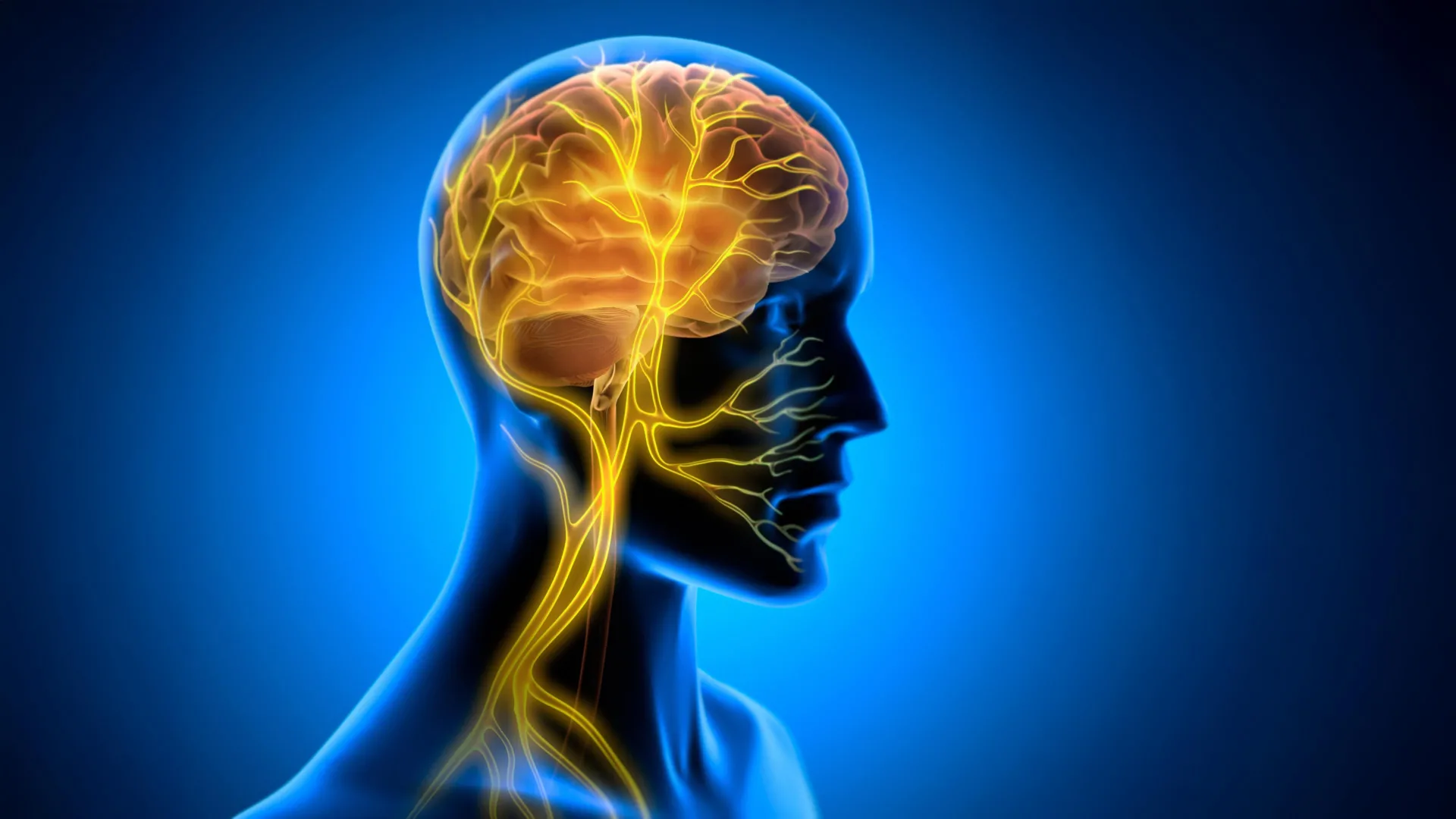
To understand the magnitude of the Tohoku University findings, one must first examine the anatomical and physiological significance of the vagus nerve. Originating in the brainstem, the vagus nerve (cranial nerve X) represents the principal component of the parasympathetic nervous system, serving as an extensive, bidirectional communication highway connecting the brain with virtually all major internal organs, including the heart, lungs, liver, and gastrointestinal tract. While it famously transmits regulatory commands downward from the brain to slow heart rate, manage digestion, and modulate inflammatory responses, a substantial majority of its nerve fibers—approximately 80%—are actually afferent. These sensory fibers carry a continuous, high-volume stream of real-time physiological data upward from the body’s viscera to the brainstem, constantly informing the central nervous system of the body’s internal state.
In recent decades, biomedical engineers and neuroscientists have sought to harness this powerful pathway through vagus nerve stimulation (VNS). Originally developed and clinically approved for the treatment of drug-resistant epilepsy and severe, treatment-refractory depression, VNS involves the surgical implantation of a small device—resembling a cardiac pacemaker—that delivers mild, intermittent electrical pulses to the left cervical branch of the vagus nerve. More recently, researchers have begun exploring non-invasive transcutaneous VNS methods, which apply electrical currents to the outer ear or the side of the neck.
Historically, neuroscientists studying the cognitive and behavioral effects of VNS have attributed its efficacy to neuromodulation. The prevailing hypothesis suggested that stimulation of the vagus nerve activates subcortical structures such as the locus coeruleus and the nucleus tractus solitarii, which in turn flood the brain with key neurotransmitters including norepinephrine, serotonin, and acetylcholine. These chemical messengers are well-known regulators of arousal, attention, and neural plasticity—the brain’s ability to reorganize itself by forming new neural connections. However, the Tohoku University research group suspected that neuromodulation via neurotransmitter release might only represent one piece of a much larger, more complex physiological puzzle.
Experimental Design and the Mechanics of Motor Learning
To test their hypothesis regarding the holistic brain-body connection, the research team, spearheaded by Professor Ko Matsui and lead author Junyu Chen, devised a rigorous experimental framework using laboratory mice. Rather than focusing on simple associative tasks, the researchers elected to study a complex, cerebellum-dependent motor learning paradigm known as the horizontal optokinetic response (HOKR).
The HOKR is an involuntary, reflexive eye movement task designed to stabilize visual images on the retina during head movement or when tracking a moving panoramic visual field. In the experimental setting, mice were placed within a specialized apparatus surrounded by moving vertical visual stripes. Over the course of training, the animals learned to coordinate precise, automatic eye movements to follow the sweeping visual stimuli—a motor skill functionally analogous to the physiological mechanisms humans employ when standing on a railway platform and visually tracking a passing train.
To manipulate the communication channel between the body and the brain during this process, the research team engineered a bespoke, miniaturized cuff electrode. This delicate device was surgically implanted and permanently secured around the left cervical vagus nerve of the subjects, allowing researchers to deliver controlled, precise electrical stimulation protocols without inducing undue physical stress or restricting the animals’ natural movements.
Crucially, the timing of the intervention became the defining variable of the study. Rather than administering VNS concurrently with the HOKR training task—the standard approach in most previous neurostimulation studies—the Tohoku University team resolved to deliver the electrical pulses strictly after the completion of each daily training session.
Chronology of Discovery: The Post-Practice Window of Opportunity
The chronological progression of the experiment yielded counterintuitive yet revealing results. During the active training phase, when the mice were undergoing the HOKR task, the application of VNS produced no immediate, observable enhancement in their motor performance. The animals exhibited standard learning curves while the electrical stimulation was absent, and their immediate tracking accuracy did not fluctuate wildly upon the delivery of post-training pulses.
However, when the researchers evaluated the subjects on subsequent days, a dramatic divergence emerged. Mice that had received VNS immediately following their training sessions demonstrated significantly superior long-term retention and motor skill execution compared to control groups that underwent training without subsequent stimulation. By Day 5 of the evaluation protocol, the VNS-treated cohort displayed robust, consolidated memory traces that translated into markedly improved visual tracking precision.
This temporal dissociation—wherein stimulation had no immediate effect on performance but drastically improved subsequent retention—provided critical insight into the mechanics of memory consolidation. In neurobiology, consolidation is the time-dependent process by which labile, newly acquired short-term memories are gradually transformed into stable, long-term structural representations within the neural network.
"The key point is that VNS was delivered only after training," noted Professor Ko Matsui, emphasizing the significance of the temporal window. "Our findings suggest that VNS may open a hidden window of opportunity for enhanced learning by making the brain environment more receptive to long-lasting change. It is not merely about accelerating execution during the act of practice, but about optimizing the biological aftermath—the crucial consolidation phase when the brain actively archives its experiences."
Unveiling the Vascular Mechanism: Rhythmic Blood Volume Oscillations
Intrigued by the profound behavioral improvements observed days after stimulation, the research team sought to identify the underlying physiological shifts occurring within the central nervous system. Specifically, they directed their investigative focus toward the cerebellar flocculus—a localized, highly specialized region of the cerebellum known to play an indispensable role in controlling vestibular-ocular reflexes and executing HOKR learning.
Employing advanced fiber photometry—an optical technique that uses fluorescent indicators to monitor neural and physiological activity deep within living tissue—the researchers measured real-time hemodynamic changes surrounding the cerebellar flocculus in response to VNS.
The optical data revealed a striking, two-phase vascular response triggered by a single round of vagus nerve stimulation. Initially, local blood volume within the targeted cerebellar microvasculature experienced a brief, transient decrease, which was swiftly followed by a pronounced, delayed rebound and elevation in blood volume. When the researchers administered repeated cycles of VNS, mimicking the post-training protocol used in the behavioral experiments, these vascular fluctuations transformed into rhythmic, oscillatory waves of blood volume changes traversing the cerebellar tissue.
Furthermore, statistical analysis demonstrated a direct correlation between the magnitude of these vascular rhythms and the degree of motor skill acquisition. Subjects that exhibited the most pronounced, robust blood volume oscillations following VNS consistently achieved the highest learning scores by the conclusion of the five-day testing period. This empirical link suggests that vagus nerve signaling does not operate purely through microscopic neurotransmitter cascades, but actively alters the macroscopic metabolic and vascular landscape of the brain, creating a hospitable physiological milieu for structural neuroplasticity.
"Our brains may be more strongly influenced by the body than we imagine," observed lead author Junyu Chen, reflecting on the broader implications of the vascular data. "By tuning the brain’s metabolic environment, including rhythmic vascular movements, we may eventually unlock capacities that would otherwise remain latent, dormant within our neural architecture."
Context, Implications, and Expert Analysis
The publication of these findings in iScience arrives at a time of paradigm shift within neurobiology. For decades, the dominant investigative lens in cognitive science has treated the brain as an isolated computational organ, analyzing synaptic transmission, ion channels, and local cortical networks while treating the rest of the body as a mere mechanical chassis designed to transport the head.
In recent years, however, the "embodied cognition" movement, coupled with discoveries regarding the gut-brain axis, the immune system’s interaction with neuroinflammation, and peripheral autonomic signaling, has forced a recalibration of this view. The Tohoku University study provides hard, mechanistic data demonstrating that peripheral physiological inputs can directly choreograph cerebral blood dynamics to facilitate memory consolidation.
Independent neuroscientists not involved in the study have praised the methodological rigor of the work, particularly the deployment of chronic, localized cuff electrodes paired with fiber photometry to track hemodynamic shifts in behaving subjects. Dr. Elena Vance, a neurophysiologist specializing in motor control and neurorehabilitation, noted that the research bridges a critical gap between clinical neuromodulation therapies and basic neuroplasticity research.
"We have known for some time that VNS accelerates stroke rehabilitation and enhances cognitive training in both animal models and human clinical trials," Dr. Vance explained. "However, the exact biological bridge between a pulse delivered to a nerve in the neck and the permanent reorganization of cerebellar circuits has remained murky. By identifying this two-phase vascular response and the resulting hemodynamic rhythms in the cerebellum, Matsui and his colleagues have provided a plausible, elegant physical mechanism. They have shown that the body can effectively ‘massage’ the brain’s metabolic state to lock in learning."
Future Directions and Clinical Horizon
While the implications of the study are profound, the research team and external analysts emphasize that translating these murine findings into human clinical applications will require substantial, methodical further investigation. Future studies will need to focus on refining stimulation protocols—determining optimal pulse frequencies, durations, and intensities—and mapping how bidirectional communication pathways modulate plasticity across different regions of the brain, such as the motor cortex and hippocampus, beyond the cerebellar flocculus.
Nevertheless, the long-term clinical and educational horizons are expansive. If researchers can successfully harness vagus nerve stimulation or non-invasive peripheral interventions to systematically open "windows of opportunity" for post-training memory consolidation, the implications for neurological rehabilitation could be transformative. Stroke survivors struggling to relearn basic motor functions, patients recovering from traumatic brain injuries, individuals dealing with degenerative movement disorders, and even students acquiring complex physical or cognitive skills could potentially benefit from targeted, post-practice physiological priming.
As science continues to dismantle the artificial boundaries separating mind and body, the work conducted at Tohoku University serves as a powerful reminder of our integrated biology. Mastery, it appears, is not merely a product of mental willpower or repetitive exertion; it is a whole-body symphony, where the silent, steady signals whispering from our internal organs help orchestrate the permanent architecture of human learning.
