
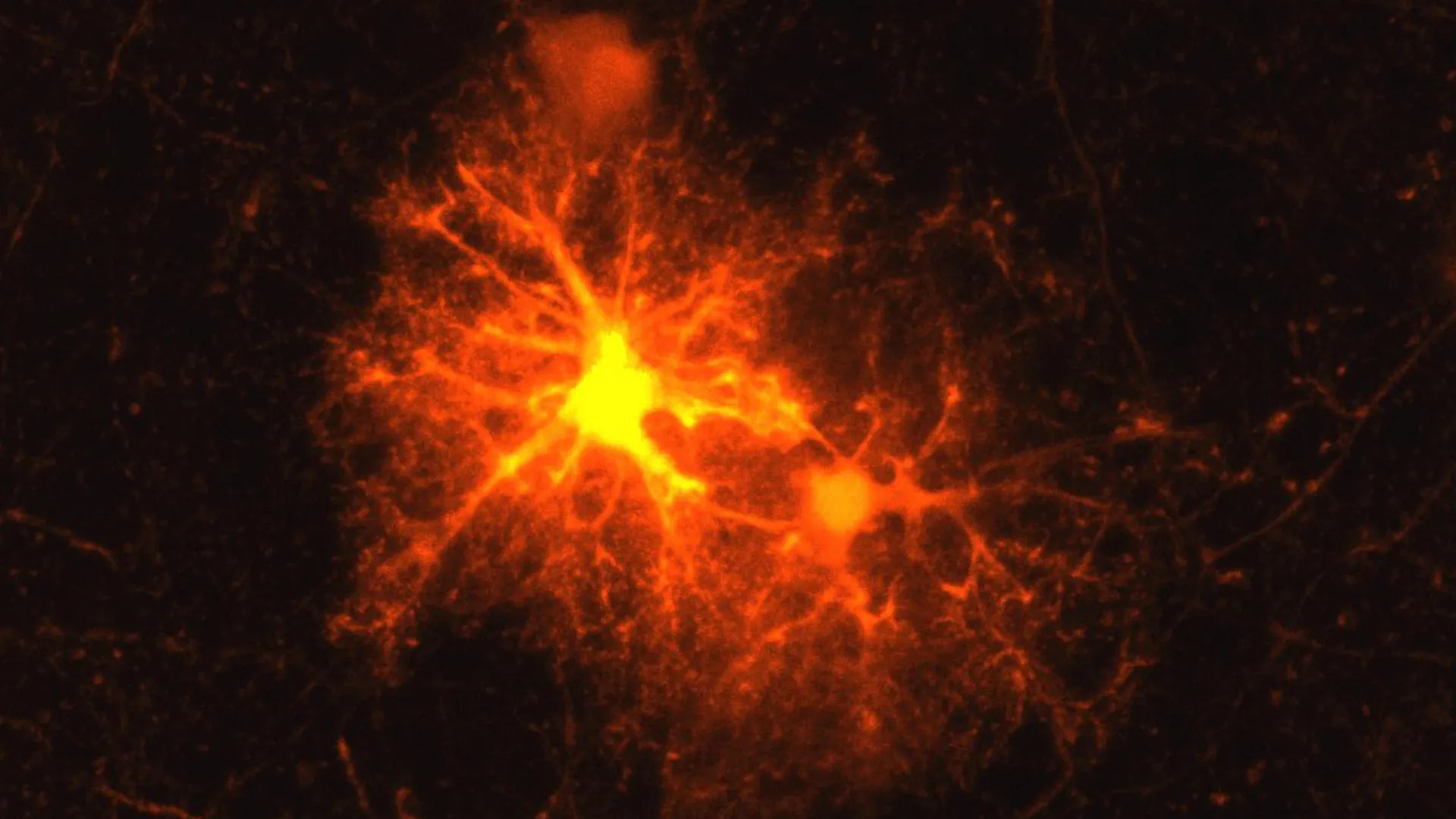
For years, neurobiologists studying laboratory models of brain trauma observed a curious and recurring phenomenon. Whenever cerebral tissue sustained localized damage—whether induced mechanically or via chemical injection—a distinct and densely clustered population of cells invariably materialized around the lesion site. To Dr. Jan Deussing, an experienced research group leader at the Max Planck Institute of Psychiatry, this cellular mobilization was a familiar sight, yet its precise biological identity remained an elusive puzzle. What were these cells, why did they selectively aggregate at the frontiers of brain wounds, and what role did they play in the organ’s intrinsic healing cascade?
These questions transformed into an ideal investigative project for Clemens Ries, a master’s degree student in biology who was wrapping up his academic training with an internship at the renowned Munich-based institute. What began as an exploratory thesis assignment evolved into a multi-year doctoral investigation that has fundamentally shifted our comprehension of neurobiology, shedding light on a surprising intersection between injury response, myelin regeneration, psychiatric vulnerability, and the body’s primary stress regulation systems.
The findings, recently published in the esteemed peer-reviewed journal Cell Reports, not only identify the mysterious responders as oligodendrocyte progenitor cells but also reveal that these precursor cells deploy a potent stress hormone—corticotropin-releasing hormone (CRH)—to coordinate the intricate timeline of tissue repair and brain development.
Identifying the Elusive Responders: Oligodendrocyte Progenitor Cells
To crack the biological mystery, Ries adopted a systematic, methodical approach utilizing an established mouse model of brain injury. Working under Deussing’s supervision, the young researcher screened molecular markers corresponding to virtually every known cell type in the central nervous system. After exhaustive testing, only one marker yielded a positive response: the signature profile of oligodendrocyte progenitor cells, commonly known as OPCs.
In a healthy, intact brain, OPCs serve as versatile precursors destined to mature into oligodendrocytes. These specialized glial cells are responsible for generating myelin, a rich lipid and protein membrane that wraps tightly around neuronal axons—the long, slender projections that facilitate electrical communication between brain cells. Functionally, myelin acts much like the synthetic insulation wrapped around copper electrical wiring. It not only accelerates the propagation of electrical impulses across neural networks—allowing signals to travel at speeds up to 100 meters per second—but also provides essential metabolic and nutritional support to the underlying axons. Without an intact myelin sheath, neuronal signaling falters, leading to functional impairment and, in advanced scenarios, axonal degeneration and cell death.
The integrity of myelin is therefore paramount to neurological health. When this protective insulation becomes degraded or destroyed—as occurs pathologically in autoimmune conditions like multiple sclerosis (MS), or traumatically through physical head injuries—the consequences can be devastating. Consequently, the rapid deployment of mechanisms capable of repairing and restoring lost myelin is one of the central imperatives of the mammalian central nervous system’s post-injury healing response.
A Timeline of Discovery: From Master’s Thesis to Breakthrough Research
The identification of OPCs as the primary cellular respondents to brain trauma formed the bedrock of Ries’s master’s thesis. However, the scientific implications of the discovery proved too profound to conclude with a standard graduate project. "The topic remained so exciting that it became my doctoral thesis," Ries reflects, charting a course of inquiry that would span several years of meticulous laboratory experimentation.
As Ries and Deussing expanded their investigations, they mapped out a clear chronology of the cellular events unfolding in the wake of a brain injury. Within hours of localized damage, OPCs residing in the surrounding parenchyma undergo a dramatic proliferative burst, accumulating heavily at the margins of the wound. The vast majority of these newly multiplied precursor cells subsequently embark on a maturation pathway, differentiating into fully functional oligodendrocytes capable of synthesizing fresh myelin to patch the breach.
Yet, alongside this expected regenerative mobilization, the researchers stumbled upon an entirely novel biological mechanism. Molecular analyses revealed that approximately one third of the OPCs localized near the damaged tissue rapidly activate the production of corticotropin-releasing hormone (CRH). Historically, CRH has been understood by endocrinologists and neurobiologists as a master regulator of the hypothalamic-pituitary-adrenal (HPA) axis, functioning as the body’s premier driver of the systemic neuroendocrine stress response. Prior to this study, the scientific community operated under the consensus that CRH production was largely restricted to specific neuronal populations within the hypothalamus and related limbic structures. The discovery that glial progenitor cells could synthesize and secrete neuropeptides such as CRH upends decades of textbook assumptions regarding cellular functional specialization in the brain.
The temporal dynamics of this CRH surge further underscored its significance. The production of the hormone by OPCs initiates remarkably fast, detectable within mere hours following the initial insult. However, this secretory burst is strictly transient, tapering off and shutting down entirely roughly three days post-injury. This narrow window of activity strongly implies that CRH acts as an urgent, early-stage molecular cue—an emergency signal designed to orchestrate the foundational phases of the neural healing cascade before stepping aside.
Decoding the Mechanism: CRH Receptor 1 and the Timing of Repair
To understand how this locally produced CRH influences tissue recovery, the research team investigated its downstream targets. The brain utilizes specific receptor proteins to bind and interpret CRH signals, principal among which are CRH receptor 1 (CRHR1) and CRH receptor 2.
The investigators discovered that CRH receptor 1 is densely expressed on a distinct population of OPCs within the vicinity of the injury, positioning these neighboring cells to intercept and respond to the CRH released by their activated peers. To test the functional necessity of this receptor-ligand interaction, the researchers evaluated injury responses in experimental models lacking functional CRHR1.
The results revealed a paradoxical outcome. In the absence of CRH receptor 1, OPCs proliferated even more aggressively following an injury than they did in control subjects. However, this hyper-proliferation did not translate into superior tissue repair. On the contrary, the initial overproduction of precursor cells proved counterproductive; ultimately, fewer mature oligodendrocytes successfully differentiated, and fewer permanent myelin repairs were established along the damaged axons.
This crucial observation provided the key to understanding the hormone’s true role. CRH does not merely stimulate or inhibit cell division; rather, it acts as a critical timing coordinator. By fine-tuning the rate of OPC proliferation and orchestrating the precise moment at which these cells transition into mature oligodendrocytes, the CRH-CRHR1 signaling axis ensures that the brain does not flood a wound site with immature cells that fail to complete their restorative mission. Proper timing, controlled by endocrine-like signaling within the local microenvironment, is essential for generating a sufficient cohort of mature myelin-producing cells to successfully restore the structural and functional integrity of damaged neural pathways.
Shaping the Developing Brain: Implications Beyond Trauma
Intrigued by the pervasive presence of CRH receptor 1 on OPCs, Ries and Deussing widened the scope of their inquiry. Because CRHR1 was identifiable on precursor cells even in the complete absence of physical injury or trauma, the researchers hypothesized that this signaling system might serve a broader, foundational purpose during normal neurodevelopment.
In the mammalian brain, the process of myelination is not completed prior to birth; rather, it is a prolonged developmental trajectory that initiates late in gestation, accelerates dramatically postnatally, and continues through adolescence well into young adulthood. Collaborating with wider research networks, the Max Planck team deployed multi-modal analytical techniques to examine myelin formation across various developmental stages in control and CRHR1-deficient mouse models.
The developmental data mirrored their injury findings in striking ways. Mice lacking CRH receptor 1 exhibited an overabundance of OPCs during early developmental epochs. Crucially, these structural deviations were not transient developmental anomalies; they persisted into adulthood, leaving permanent architectural alterations across the brain’s white matter tracts.
Histological examination of adult brains from these models revealed pronounced structural changes in myelination, characterized notably by significantly thicker myelin sheaths enveloping thin axons. These insights demonstrate that the CRH-CRHR1 axis on OPCs is a dual-purpose biological machinery: it is recruited during adulthood to manage the acute exigencies of tissue repair, yet it is equally indispensable during embryogenesis and juvenile maturation for establishing the baseline architecture of the central nervous system.
While the source of CRH during an acute injury is clearly the reactive OPCs themselves, the physiological origin of CRH during normal brain development presented a secondary biological puzzle. How is the signaling pathway activated when there is no tissue trauma to trigger local glial production?
Synthesizing current neurobiological data, the researchers propose a compelling hypothesis centered on neuronal activity. During normal maturation, developing neurons are known to synthesize and secrete neuropeptides. The team posits that juvenile neurons release CRH into the extracellular milieu, where it diffuses to interact with CRHR1 on neighboring OPCs, thereby governing their developmental multiplication and directing their eventual maturation into myelin-forming oligodendrocytes.
Broader Horizons: Connecting the CRH System to Stress and Mental Health
Because neurons are well-documented to ramp up CRH release in response to physiological and psychological stressors—and given that early childhood adversity and chronic stress are universally recognized epidemiological risk factors for the onset of psychiatric disorders such as major depressive disorder, anxiety, and post-traumatic stress disorder—these findings open intriguing new avenues for neuropsychiatric research.
If glial-derived and neuron-derived CRH signaling pathways actively shape the structural composition of white matter tracts during formative developmental windows, perturbations in this system could have profound implications for mental health. Chronic exposure to stress hormones during critical developmental epochs might subtly alter OPC proliferation and myelination trajectories, predisposing individuals to structural neural vulnerabilities later in life.
"Our current findings suggest that in stress-associated psychiatric disorders such as depression, the CRH system in OPCs may play a greater role than previously known," Dr. Deussing speculates, framing the study within a broader clinical context.
While speculative, this hypothesis shifts the prevailing paradigm in psychiatry and neurobiology. Traditionally, research into stress-related mood disorders has focused almost exclusively on neuronal circuits, synaptic neurotransmission, and endocrine feedback loops. The revelation that glial progenitor cells—and the myelin they produce—are active participants in CRH-mediated stress responses suggests that structural alterations in white matter may be an under-appreciated component of psychiatric pathology.
As research moves forward, the scientific community will look to build upon these foundational insights. Future studies will need to determine whether the mechanisms observed in murine models translate faithfully to the human central nervous system and whether targeted pharmacological modulation of CRH signaling in OPCs can be harnessed therapeutically. Should subsequent investigations validate these connections, deciphering how CRH influences myelin formation and brain plasticity could eventually pave the way for entirely novel therapeutic strategies aimed at treating neurodegenerative conditions, traumatic brain injuries, and severe stress-associated psychiatric disorders.
