
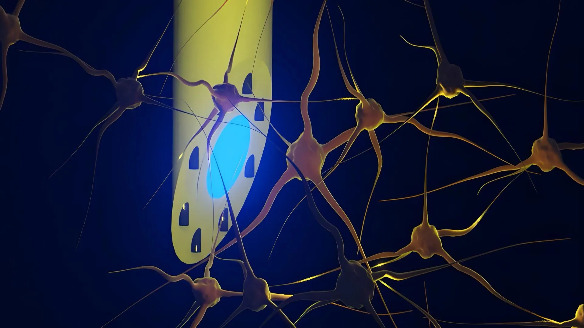
An international coalition of biomedical engineers and neuroscientists has unveiled a breakthrough neural interface device that promises to fundamentally change how researchers investigate complex neurological disorders. Developed through a collaborative effort involving the Technical University of Denmark (DTU), the University of Copenhagen, and University College London (UCL), the newly engineered implant—known as the microfluidic Axialtrode, or mAxialtrode—combines optical capabilities, microfluidic channels, and electrical recording components into a single, highly flexible probe thinner than a human hair. Published in the peer-reviewed journal Advanced Science, this pioneering technology bridges a long-standing technological gap in neurophysiology by allowing scientists to simultaneously monitor electrical brain activity, deliver precise micro-doses of therapeutic or pharmacological agents, and apply localized optical stimulation across multiple distinct depths of living neural tissue.
Main Facts and the Core Innovation
At its core, the mAxialtrode addresses a critical limitation inherent in traditional brain-computer interfaces and neuroscientific research tools: the trade-off between invasiveness and functionality. For decades, electrophysiologists studying animal models of epilepsy, memory formation, and cognitive decision-making have been forced to rely on rigid silicon arrays, heavy metallic electrodes, or conventional, flat-ended optical fibers. These legacy tools typically interact with brain tissue only at their extreme distal tips, severely restricting researchers to recording or stimulating a single localized layer of the brain at any given moment.
Furthermore, because many complex neurological functions—and pathological events such as epileptic seizures—depend on rapid, synchronized communication across multiple interconnected brain layers and deep subcortical structures, studying these processes required the simultaneous surgical implantation of numerous independent devices. This multi-device approach inevitably increases tissue trauma, elevates the risk of localized chronic inflammation, and complicates experimental protocols.
The mAxialtrode resolves these complications by integrating multiple functionalities into a single polymer-based filament measuring less than half a millimeter in diameter. Manufactured through an advanced thermal drawing process—where a macroscopic polymer preform is heated and pulled under precise mechanical tension—the final strand features a central light-conducting core surrounded by eight microscopic fluidic channels and ultra-thin embedded metal wires. This unique architecture enables researchers to introduce light for optogenetic activation, administer pharmaceutical compounds, and record electrical neural signals concurrently at multiple functional points along the entire length of a single implanted probe.
Chronology and Development Timeline
The genesis of the mAxialtrode dates back several years to foundational discussions within the biomedical engineering laboratories at DTU, where Postdoc Kunyang Sui and Associate Professor Christos Markos sought to overcome the mechanical and functional limitations of conventional neural probes. Recognizing that conventional glass and hard silicon fibers create a severe stiffness mismatch with soft biological neural tissue—often prompting glial scarring and immune reactions that degrade signal quality over time—the team pivoted toward flexible polymers.
By optimizing the thermal drawing technique, Sui and Markos successfully engineered a fiber featuring an innovative, specially angled tip and distributed microfluidic architecture. Following initial prototyping and benchtop validation, the research team expanded its collaborative network to include neurophysiologists and clinical epilepsy researchers, notably Associate Professor Rune W. Berg of the University of Copenhagen and Associate Professor Rob C. Wykes of University College London.
This multidisciplinary partnership enabled the team to transition the technology from in vitro testing to rigorous in vivo validation. Over the past year, the mAxialtrode was surgically implanted into living murine (mouse) models. These preclinical trials were designed to test the probe’s mechanical durability, biocompatibility, and functional efficacy in deep-brain structures. The successful in vivo demonstrations—recording high-fidelity electrical signals from both the superficial cerebral cortex and the deep hippocampus while simultaneously injecting chemical agents and delivering multi-color light stimulation—marked a major milestone, proving that the device could function seamlessly within a living, moving mammalian brain without causing apparent physiological distress to the subjects.
Supporting Data and Technical Specifications
To fully appreciate the engineering achievement represented by the mAxialtrode, it is necessary to examine the physical and operational parameters that differentiate it from standard neuroscientific hardware. Traditional optical fibers, widely utilized in optogenetics since the early 2000s, are typically rigid cylinders of glass or hard plastic. When inserted into the rodent brain, these fibers exert mechanical stress, leading to gradual encapsulation by glial cells and a subsequent loss of recording fidelity within weeks.
In contrast, the mAxialtrode is manufactured from soft, biocompatible thermoplastic polymers that closely match the mechanical compliance of soft neural tissue. This flexibility allows the implant to micro-move in tandem with natural cerebral pulsations, drastically reducing chronic inflammatory responses. Structurally, the fiber houses:
- A central optical waveguide capable of transmitting both red and blue light wavelengths deep into subcortical regions.
- Eight independent microfluidic capillary channels capable of transporting liquid pharmacological solutions.
- Integrated metallic micro-wires capable of picking up minute local field potentials and action potentials from surrounding neurons.
During in vivo testing, researchers demonstrated the device’s precise fluidic delivery capabilities by injecting distinct substances at separate tissue depths spaced nearly three millimeters apart along a single insertion tract. Simultaneously, the optical components successfully activated target neural populations via light-gated ion channels, while the embedded electrodes captured real-time electrophysiological responses across different cortical and hippocampal strata. The entire assembly was light enough to be carried by freely moving mice, ensuring that behavioral and cognitive experiments could proceed without physical impediment.
Official Responses and Perspectives from the Research Team
The development of the mAxialtrode has generated considerable enthusiasm within the European neurotechnology community, eliciting detailed reflections from the principal investigators and their institutional collaborators.
"Most current brain implants are based on hard materials such as silicon, which can irritate the brain and trigger inflammatory reactions in the tissue," explained Dr. Kunyang Sui, co-creator of the device alongside Associate Professor Christos Markos. "The new implant differs in that it is made of soft, plastic-like optical fibers and has a specially angled tip that makes it smaller and reduces the damage caused when it is placed in the brain. One of the main advantages is that several capabilities can be combined within a single implant, allowing researchers to perform more precise experiments while reducing the need for multiple devices."
While the scientific utility for basic neuroscience research is immediately apparent, the team emphasizes caution regarding immediate clinical applications. Dr. Sui and his colleagues stress that translational medicine requires a rigorous, phased approach.
"The technology is still far from routine clinical use," Sui noted, adding that extensive preclinical safety testing, long-term biocompatibility studies, scale-up manufacturing validation, and comprehensive regulatory reviews by bodies such as the European Medicines Agency (EMA) and the U.S. Food and Drug Administration (FDA) will be mandatory before human trials can even be considered.
Associate Professor Rune W. Berg of the University of Copenhagen, who contributed expertise in neural circuit analysis and motor control, underscored the importance of the device in decoding complex network dynamics. "Understanding how neural information flows dynamically through layered brain structures has always been hampered by our instrumentation," Berg stated. "With the mAxialtrode, we can observe and manipulate these circuits with a spatial and temporal granularity that was previously unachievable using isolated methods."
Similarly, Associate Professor Rob C. Wykes of University College London highlighted the relevance of the technology to pathological states such as drug-resistant epilepsy. "Epileptic seizures do not originate in a vacuum; they recruit vast, distributed networks spanning multiple brain regions," Wykes explained. "Tools that can simultaneously monitor these networks, deliver anti-epileptic drugs precisely where and when they are needed, and apply electrical or optogenetic neuromodulation represent an exciting frontier in potential therapeutic intervention."
Broader Impact, Future Implications, and Clinical Horizons
The implications of the mAxialtrode extend well beyond the immediate confines of academic neurobiology laboratories. As the global prevalence of neurological disorders—including epilepsy, Parkinson’s disease, Alzheimer’s disease, and major depressive disorder—continues to rise alongside aging populations, the demand for more sophisticated, targeted neurotechnologies has never been more urgent.
Current clinical neurostimulation devices, such as deep brain stimulation (DBS) systems used for Parkinson’s disease and severe epilepsy, rely predominantly on electrical impulses delivered via rigid metallic leads. While these devices have successfully alleviated symptoms for hundreds of thousands of patients worldwide, they suffer from a lack of chemical specificity and often stimulate healthy adjacent tissue, leading to unwanted side effects.
The convergence of optical stimulation, localized fluidic drug delivery, and high-density electrical recording embodied by the mAxialtrode points toward a sophisticated, closed-loop therapeutic paradigm. In a hypothetical future clinical scenario, a bio-integrated variant of such an implant could continuously monitor local electrocorticographic activity to detect the earliest aberrant neural firing patterns associated with an impending epileptic seizure. Upon detection, the system could automatically deploy a dual-action response: instantly releasing a minute micro-dose of an anti-seizure medication directly into the seizure focus via the microfluidic channels, while simultaneously applying localized light or electrical stimulation to interrupt the pathological circuit before a clinical seizure can manifest.
Such closed-loop, multimodal therapies could drastically reduce the systemic side effects associated with oral medications, which flood the entire bloodstream with pharmacologically active compounds that affect non-target organs. Furthermore, the soft, flexible nature of polymer-based fibers suggests that future neural implants could remain implanted safely for decades without inducing the chronic tissue encapsulation that currently limits the lifespan of traditional metallic brain electrodes.
Commercialization and Next Steps
Recognizing the immense translational potential of their invention, the research consortium is taking proactive measures to secure the intellectual property rights associated with the mAxialtrode. The team is currently working to patent the core manufacturing processes, architectural designs, and integrated functional methodologies behind the brain electrode.
Concurrently, the researchers are engaging with academic technology transfer offices and industrial partners to map out a commercialization pathway. This phase involves refining the mass-production of the polymer fibers to ensure high batch-to-batch reproducibility, standardizing the integration of micro-wires and fluidic connectors, and designing automated packaging solutions suitable for sterile medical manufacturing.
As patent applications proceed and discussions regarding clinical translation gain momentum, the mAxialtrode stands as a testament to the power of interdisciplinary collaboration. By fusing advanced materials science, polymer chemistry, microfluidics, and neurophysiology, DTU, the University of Copenhagen, and University College London have provided the scientific community with an unprecedented instrument for decoding the brain—bringing researchers one step closer to unravelling the most intricate mysteries of human cognition and pathology.
