
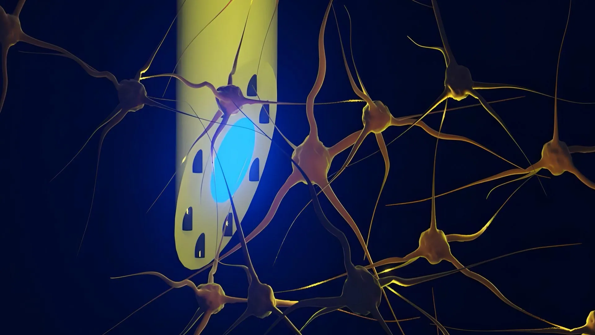
An international consortium of researchers has unveiled a revolutionary brain implant that combines electrical recording, optical stimulation, and targeted drug delivery into a single, needle-thin device. Developed by a multidisciplinary team from the Technical University of Denmark (DTU), the University of Copenhagen, and University College London, the invention—formally named the microfluidic Axialtrode (mAxialtrode)—addresses longstanding engineering limitations in neuroscience. Measuring less than half a millimeter in diameter, this flexible, polymer-based fiber allows scientists to interact with multiple layers of brain tissue simultaneously. Published in the peer-reviewed journal Advanced Science, the breakthrough promises to transform how researchers investigate complex neurological disorders such as epilepsy, Alzheimer’s disease, and major depressive disorders, while laying the groundwork for a new generation of closed-loop clinical therapies.
The Evolution of Neural Interfaces: A Historical Chronology
To understand the significance of the mAxialtrode, one must examine the historical trajectory of brain-computer interfaces (BCIs) and neural probes. For decades, neuroscience has relied on rigid silicon arrays, such as the Utah array, or metal microelectrodes to record electrical spikes from individual neurons. While these hard implants successfully advanced our understanding of single-unit electrophysiology, they presented a severe biological drawback: mechanical mismatch. The human brain is a soft, gelatinous organ that shifts continuously within the cranial vault. Rigid silicon probes remain stationary, causing chronic friction, micro-motion injuries, and subsequent glial scar tissue formation. This inflammatory response often degrades signal quality over weeks or months, limiting the lifespan of the implant.
In the early 2000s, the advent of optogenetics revolutionized neuroscience by allowing researchers to genetically modify neurons to respond to specific wavelengths of light. This technique required the integration of optical fibers alongside electrical electrodes. However, conventional optical fibers were typically made of rigid glass or thick plastic with flat-ended distal tips. These legacy fibers could only deliver light or record data at a single, isolated terminus. Consequently, mapping neural circuits that spanned multiple cortical layers or extended from the superficial cortex down to deep structures like the hippocampus required the implantation of multiple, bulky devices—compounding tissue trauma and complicating experimental setups.
Recognizing these limitations, Postdoc Kunyang Sui and Associate Professor Christos Markos at DTU spearheaded an initiative to reinvent the optical fiber. Over several years of collaborative design, the research team engineered a multi-functional fiber that could bridge the gap between optical stimulation, electrophysiological recording, and pharmacology. By utilizing advanced thermal drawing techniques—similar to pulling glass or polymer into microscopic threads under controlled heat—the team successfully manufactured a sub-millimeter strand containing a central light-conducting core, eight independent microfluidic channels, and integrated micro-wires. The resulting mAxialtrode represents a paradigm shift from single-point stimulation to distributed, multi-modal brain interrogation.
Anatomy of the mAxialtrode: Engineering and Specifications
The physical architecture of the mAxialtrode distinguishes it starkly from traditional neural probes. Fabricated from biocompatible polymers rather than brittle silicon or glass, the implant boasts a Young’s modulus closely matching that of native brain tissue. This flexibility ensures that the fiber moves synchronously with the brain, drastically mitigating the inflammatory responses and glial scarring that plague conventional silicon arrays.
The manufacturing process itself is a marvel of materials science. The fiber originates as a macroscopic polymer preform containing precisely positioned geometric layouts. When heated and drawn down under high tension, the macroscopic structure scales down uniformly to a microscopic filament measuring less than 0.5 millimeters across. Running through the exact center of this flexible filament is an optical core capable of guiding light deep into subcortical structures without significant attenuation.
Surrounding this optical core are eight microscopic fluidic channels. These channels serve a dual purpose: they can transport minute quantities of liquid medications, neuromodulators, or chemical tracers directly to targeted cellular populations, and they can house ultra-thin metallic wires dedicated to recording electrical potentials. By angling the distal tip of the fiber during fabrication, the researchers created multiple functional emission and recording points along the longitudinal axis of the implant. This spatial distribution allows the mAxialtrode to simultaneously interface with different cortical layers and deep brain structures separated by distances of up to three millimeters, all through a single insertion tract.
In Vivo Validation and Experimental Results
The theoretical promise of the mAxialtrode has now been substantiated by rigorous empirical testing. In a series of in vivo experiments conducted in close collaboration with neurophysiologist Associate Professor Rune W. Berg of the University of Copenhagen and circuit specialist Associate Professor Rob C. Wykes of University College London, the device was implanted into the brains of living mice.
The animal subjects were fitted with lightweight connections linking the mAxialtrode to external diode lasers, electrophysiological recording amplifiers, and programmable micro-pumps. The subjects demonstrated remarkable tolerance to the implant, carrying the peripheral connections and the flexible fiber without exhibiting behavioral distress or motor impairment.
During the trials, the research team successfully administered both blue and red light through the central core to execute optogenetic activation of targeted neuronal populations. Concurrently, the integrated micro-wires recorded high-fidelity local field potentials and action potentials from both superficial cortical layers and deeper structures, such as the hippocampus. Furthermore, the microfluidic channels successfully delivered distinct pharmacological agents at discrete depths along the single insertion track. This multi-modal capability confirmed that researchers could observe neural circuit dynamics, apply optical stimulation, and alter chemical environments simultaneously at precise spatial coordinates.
Implications for Epilepsy Research and Therapeutics
While the mAxialtrode is currently positioned as a foundational research instrument, its long-term clinical implications are profound, particularly for the treatment of drug-resistant epilepsy. Epilepsy is characterized by aberrant, hypersynchronous electrical discharges originating from focal brain regions or propagating through established neural networks. Current neurosurgical interventions often involve the permanent surgical resection of the epileptogenic zone or the implantation of crude neurostimulators that deliver continuous electrical pulses, frequently causing unwanted side effects.
The mAxialtrode offers a sophisticated blueprint for closed-loop, multimodal epilepsy therapies. By combining real-time electrical recording with targeted pharmacological delivery and localized photostimulation, future clinical devices could detect the precursor signatures of an impending seizure, automatically release an anti-epileptic drug precisely at the focal point, and apply stabilizing electrical or optical modulation to suppress the abnormal discharge. Because the implant is soft and causes minimal tissue irritation, it could theoretically remain stable within a human patient’s brain for extended periods, reducing the need for revision surgeries.
Expert Perspectives and Official Responses
The collaborative nature of the project has drawn praise from across the European scientific community. Researchers emphasize that the integration of optical, electrical, and chemical modalities into a single strand solves a persistent bottleneck in systems neuroscience.
"Most current brain implants are based on hard materials such as silicon, which can irritate the brain and trigger inflammatory reactions in the tissue," explained Kunyang Sui, co-inventor of the mAxialtrode. "The new implant differs in that it is made of soft, plastic-like optical fibers and has a specially angled tip that makes it smaller and reduces the damage caused when it is placed in the brain."
Sui and his colleagues are careful to contextualize the current state of the technology. Despite the successful animal trials, the transition from an experimental laboratory tool to a certified medical device is a rigorous, multi-year undertaking. Extensive biocompatibility testing, chronic safety evaluations in larger animal models, and comprehensive reviews by regulatory bodies such as the European Medicines Agency (EMA) and the U.S. Food and Drug Administration (FDA) must be completed before human clinical trials can be considered.
Path Forward: Commercialization and Clinical Translation
Following the publication of their findings in Advanced Science, the DTU-led consortium is actively pursuing intellectual property protection. The research team has initiated patent filings for the mAxialtrode manufacturing process and design architecture. Concurrently, the scientists are engaging with medical technology transfer offices and industry partners to explore pathways for commercial scalability.
The immediate next steps involve refining the fluidic delivery mechanisms to ensure absolute volumetric precision and scaling up the manufacturing yield of the polymer fibers. As the scientific community moves deeper into an era defined by precision medicine and brain-computer interfacing, inventions like the mAxialtrode highlight the critical convergence of materials science, neurobiology, and microfluidics. By providing a gentler, more versatile window into the mind, this needle-thin fiber brings researchers one step closer to decoding the brain’s most intricate language and alleviating the burden of debilitating neurological diseases.
