
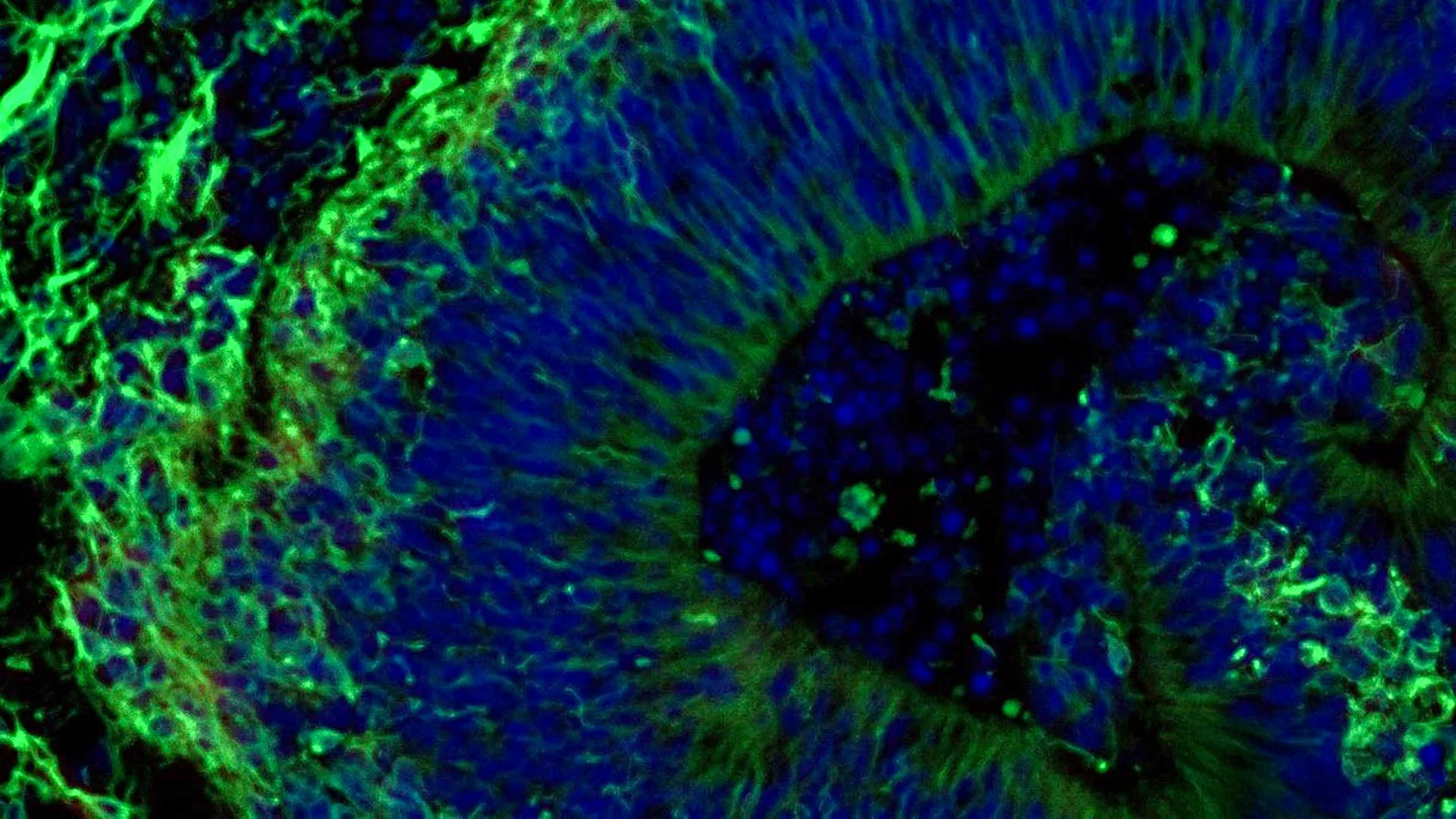
In a significant breakthrough for neurological research, scientists at Johns Hopkins Medicine have successfully utilized laboratory-grown miniature brain models—known as organoids—to predict how individual Alzheimer’s disease patients might respond to specific psychiatric medications. This milestone study, published in Alzheimer’s & Dementia: The Journal of the Alzheimer’s Association, bridges a critical gap in neuropharmacology, offering a potential path forward for tailoring treatments to the unique molecular profiles of those suffering from the most common form of dementia.
Alzheimer’s disease currently impacts more than 7 million Americans, a staggering demographic figure that continues to escalate alongside global aging trends. While the neurodegenerative condition is universally characterized by cognitive decline and memory loss, it also triggers severe neuropsychiatric symptoms—including chronic anxiety, profound depression, and debilitating agitation. These psychiatric manifestations affect nearly all diagnosed individuals at some point during the disease’s progression. To manage these symptoms, clinicians routinely prescribe selective serotonin reuptake inhibitors (SSRIs), a class of antidepressants. However, patient responses to these medications vary wildly, often leaving physicians to rely on a frustrating trial-and-error approach that can exacerbate patient distress and delay effective symptom management. The latest findings from Johns Hopkins aim to dismantle this paradigm by leveraging patient-specific tissue models to forecast drug efficacy before a single pill is prescribed.
Chronology and Evolution of Brain Organoid Technology
The realization of patient-derived brain organoids represents decades of rapid advancements in stem cell biology and tissue engineering. The foundational timeline of this research traces back to early breakthroughs in reprogramming somatic cells into induced pluripotent stem cells (iPSCs), a technique that earned a Nobel Prize and revolutionized regenerative medicine. By taking routine blood samples from human donors, researchers gained the unprecedented ability to revert mature cells back into an embryonic-like stem cell state.
Building upon this capability, the Johns Hopkins research team—led by Vasiliki Machairaki, Ph.D., associate professor of genetic medicine at the Johns Hopkins University School of Medicine—utilized blood samples collected with full consent from individuals with Alzheimer’s disease. These samples were sourced via the NIH-funded Johns Hopkins Alzheimer’s Disease Research Center.
Once the cells were reprogrammed into iPSCs, the scientists directed their development specifically toward the hindbrain, a vital anatomical region located at the back of the skull responsible for regulating core autonomic functions such as breathing, sleep cycles, and heart rate. Through careful biochemical signaling, these cells organized themselves into pea-sized clusters of functional brain tissue. Containing specialized neurons capable of producing the neurotransmitter serotonin, these hindbrain organoids effectively served as living, micro-scale avatars of the donors.
Encompassing hundreds of individual organoids representing both Alzheimer’s patients and healthy control participants, this project stands as one of the most extensive brain organoid studies conducted to date within the field of dementia research. The creation of these models allows scientists to bypass the ethical and logistical impossibilities of directly sampling living human brain tissue, creating a scalable platform for high-throughput pharmacological testing.
Unlocking Molecular Differences and Drug Responses
Upon establishing the hindbrain organoids, the research team conducted deep molecular profiling to evaluate how accurately the lab-grown tissue mirrored the pathology of living Alzheimer’s brains. The comparisons revealed striking distinctions. Organoids grown from the cells of Alzheimer’s patients exhibited fundamental alterations in proteins tied directly to neuroinflammation, synaptic communication, and established disease-related pathways, distinguishing them clearly from organoids derived from healthy individuals.
With the disease phenotype successfully replicated in vitro, the investigators introduced escitalopram oxalate, a widely prescribed SSRI antidepressant, to the culture environment. The results demonstrated a clear divergence in tissue behavior. In a subset of patient-derived organoids, the medication successfully upregulated proteins involved in serotonin signaling and enhanced intercellular communication—precisely the biological pathways that antidepressants are engineered to modulate. Conversely, other patient organoids exhibited negligible or zero molecular response to the drug.
This variance provides a mechanistic explanation for why clinical responses to SSRIs are so unpredictable among human patients. According to Machairaki, observing these distinct molecular signatures in a controlled laboratory setting paves the way for categorizing patients into specific subgroups. By identifying which underlying biological mechanisms correlate with a positive pharmacological response, clinicians may eventually bypass ineffective therapies, drastically shortening the timeline to relief for vulnerable patients.
Extracellular Vesicles as Novel Liquid Biopsies
Beyond the cellular analysis of the organoids themselves, the Johns Hopkins team made a secondary discovery that could fundamentally transform Alzheimer’s diagnostics: the analysis of extracellular vesicles. These tiny, fluid-filled particles are naturally released by the organoids into their surrounding environment, carrying a rich cargo of cellular information, including proteins, lipids, and nucleic acids.
Before and after administering escitalopram oxalate, the scientists isolated and examined the proteins contained within these secreted vesicles from both Alzheimer’s and control organoids. The findings were revealing. Vesicles derived from Alzheimer’s organoids demonstrated marked reductions in essential signaling proteins—specifically RAB3A, NSF, and ATCAY—which are critical for normal neurotransmission and synaptic maintenance.
Following treatment with the antidepressant, the protein profiles within the vesicles shifted, mirroring the molecular changes observed inside the tissue itself. Once again, a distinct polarization emerged: certain samples showed robust molecular shifts in response to the drug, while others remained static.
This correlation suggests that extracellular vesicles could serve as non-invasive biomarkers, or "liquid biopsies," for Alzheimer’s disease. In a clinical setting, drawing a patient’s blood or cerebrospinal fluid to analyze circulating vesicles could theoretically allow physicians to stage the disease, identify specific molecular subtypes, and predict treatment responsiveness without requiring invasive tissue extraction.
Official Responses and Collaborative Support
The implications of this study extend well beyond individual symptom management, touching upon the broader philosophy of precision medicine in neurology. The research underscores the growing shift away from generalized treatment guidelines toward individualized care models tailored to a patient’s unique genetic and molecular architecture.
"Our study suggests that large-scale, patient-derived brain organoids and the vesicles they secrete can help us stage Alzheimer’s disease, investigate the mechanisms that drive it and assess how patient subgroups may respond to different treatments," stated Dr. Machairaki. She emphasized that while the findings are promising, the project remains an essential early step toward realizing fully personalized neurological care.
The interdisciplinary nature of the study brought together leading experts across multiple institutions. Alongside Machairaki, the research team included Rachel Boyd, Daiyun Dong, Ram Sagar, Waqar Ahmed, Xenia Androni, Paul Rosenberg, Constantine Lyketsos, and Kenneth Witwer from Johns Hopkins; Anton Iliuk from Tymora Analytical Operations; and Anton Porsteinsson from the University of Rochester School of Medicine and Dentistry.
Financial backing for the ambitious project was made possible through robust public and private partnerships. Primary funding was provided by the National Institutes of Health under grants including T32 AG058527, R01AG052510, P30AG066507, 1RF1AG083801, AGR01054771, AGR01050515, AGR01046543, and AGR01071522. Additional support was contributed by the Paul G. Allen Frontiers Foundation and the Richman Family Precision Medicine Center of Excellence in Alzheimer’s Disease at The Johns Hopkins University. According to institutional disclosures, none of the study’s authors reported any related conflicts of interest.
Broader Impact and Future Directions
As the medical community grapples with the rising global prevalence of dementia, innovations that accelerate drug development and refine patient stratification are urgently required. Traditional clinical trials for Alzheimer’s therapies frequently face hurdles due to the extreme heterogeneity of the patient population; a drug that fails in a broad, generalized trial might have succeeded if targeted specifically to a responsive biological subgroup.
Looking forward, Machairaki and her colleagues are already working on the next generation of brain organoid models. Future iterations of the technology aim to incorporate human immune cells and vascular-like capillary networks, mimicking the complex microenvironment of the living human brain more accurately than current models allow.
If these advancements successfully bridge the gap between bench science and clinical application, the potential impact on healthcare systems could be profound. By utilizing patient-derived organoids to screen psychiatric medications and relying on extracellular vesicle liquid biopsies to monitor disease progression, neuroscientists are moving closer to a future where Alzheimer’s care is no longer managed through generalized guesswork, but guided with precision medicine.
