
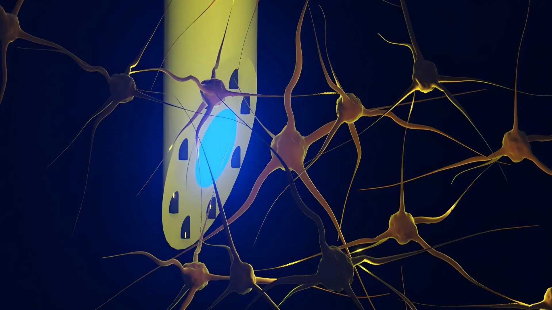
An international consortium of neuroscientists and materials engineers has unveiled a breakthrough medical device that promises to fundamentally reshape how researchers map complex neural circuits and, eventually, treat debilitating neurological disorders such as epilepsy. Published recently in the peer-reviewed journal Advanced Science, the innovation centers on a needle-thin, multi-functional brain electrode designated as the microfluidic Axialtrode, or mAxialtrode. Developed through a collaborative effort involving researchers from the Technical University of Denmark (DTU), the University of Copenhagen, and University College London (UCL), among other institutions, the implant combines electrical recording, fluid delivery, and optical stimulation capabilities into a single, highly flexible polymer strand. By integrating these previously disparate functions into a profile measuring less than half a millimeter across, the device overcomes several longstanding limitations of conventional brain-computer interfaces and rigid silicon probes.
Background Context of Neural Interface Technology
To appreciate the significance of the mAxialtrode, one must examine the trajectory of neuroengineering over the past several decades. For years, the study of brain function has relied heavily on rigid silicon-based microelectrode arrays or traditional glass and plastic optical fibers. While these legacy tools have driven monumental discoveries in neuroscience—such as the mapping of specific neural pathways and the advent of optogenetics, which allows scientists to control genetically modified neurons using light—they suffer from inherent physical constraints. Traditional optical fibers, for instance, typically interact with neural tissue exclusively at their distal tips, or "noses." This terminal restriction means that a conventional fiber can generally stimulate or monitor only a single focal point or brain layer at a time.
Furthermore, the mechanical mismatch between rigid materials like silicon or glass and the delicate, gelatinous consistency of living brain tissue has always presented a major biocompatibility hurdle. When stiff implants reside within the cranium, micro-movements caused by vascular pulsation and respiration can lead to chronic friction. This mechanical irritation routinely triggers immune responses, leading to glial scarring and tissue inflammation that eventually insulates the electrode, degrades signal quality, and limits the longevity of the recording device. In clinical settings, these inflammatory processes have restricted the duration and reliability of long-term neural monitoring, creating an urgent demand for softer, multi-modal alternatives that can integrate seamlessly with biological tissue.
Chronology and Development of the mAxialtrode
The creation of the mAxialtrode is the culmination of years of iterative materials science and neurophysiological research. The foundational concept was originally conceived by DTU Postdoc Kunyang Sui and Associate Professor Christos Markos, who sought to bridge the gap between fluidics, photonics, and electrophysiology. Recognizing that modern neuroscience requires simultaneous multi-site interventions, the team engineered a fabrication process adapted from advanced fiber-drawing techniques.
The manufacturing workflow begins with the assembly of a macro-scale polymer preform. This bulk structure contains a precisely arranged architecture of specialized components, including a central light-conducting core, eight microscopic channels designed for fluid transport, and hollow conduits intended to house ultra-thin metal wires for electrical recording. Once assembled, the preform is heated to a controlled softening point and mechanically drawn down into an exceptionally fine, continuous fiber—a process conceptually akin to producing a high-precision strand of glass or sugar, but executed with microscopic tolerances.
Following the successful benchtop fabrication and material characterization of the fibers, the DTU team partnered with neurophysiologists, including Associate Professor Rune W. Berg of the University of Copenhagen and Associate Professor Rob C. Wykes of University College London. This phase of the chronology shifted from material physics to in vivo validation. The researchers surgically implanted the mAxialtrode into living murine models to evaluate its mechanical stability, biocompatibility, and functional performance under physiological conditions.
In these live animal experiments, the implant was coupled with miniature external fluid pumps, multi-channel recording hardware, and laser or LED light sources. The trials successfully demonstrated that the mAxialtrode could emit both blue and red light to trigger optogenetic responses, record high-fidelity electrical spikes across both superficial cortical layers and deeper subcortical structures like the hippocampus, and inject pharmacological agents at distinct depths spaced nearly three millimeters apart. Crucially, the subjects carried the lightweight assembly without displaying signs of behavioral distress or physical impediment, marking a critical milestone in the device’s development timeline.
Supporting Data and Technical Specifications
The technical metrics achieved by the mAxialtrode highlight a significant leap forward in micro-scale engineering. Measuring less than 0.5 millimeters in total diameter, the implant achieves a high degree of miniaturization while packing multiple functional modalities into its cross-section.
Key technical parameters of the mAxialtrode system include:
- Diameter: Sub-millimeter profile (less than 0.5 mm across), significantly reducing tissue displacement compared to conventional bundled arrays.
- Material Composition: Soft, flexible, plastic-like optical polymers that closely match the elastic modulus of native brain tissue, mitigating chronic inflammatory responses.
- Functional Channels: A central optical core surrounded by eight microfluidic channels capable of transporting liquids and housing ultra-thin electrical recording wires.
- Spatial Resolution: Capable of drug delivery and signal acquisition across multiple functional points along the longitudinal axis of a single implant, with fluid injection sites spaced up to approximately three millimeters apart.
- Multi-Modal Stimulation: Proven compatibility with concurrent optical activation (using red and blue wavelengths) and electrical monitoring across the cerebral cortex and hippocampus.
Official Responses and Expert Analysis
The collaborative nature of the research brought together specialists from distinct disciplines, resulting in a multifaceted evaluation of the device’s potential. According to Kunyang Sui, the primary advantage of the new design lies in consolidation and tissue preservation. "Most current brain implants are based on hard materials such as silicon, which can irritate the brain and trigger inflammatory reactions in the tissue," Sui explained. "The new implant differs in that it is made of soft, plastic-like optical fibers and has a specially angled tip that makes it smaller and reduces the damage caused when it is placed in the brain."
Despite the enthusiasm surrounding the publication in Advanced Science, the research team maintains a rigorous and cautious perspective regarding the timeline for human application. Sui and his colleagues emphasize that the technology remains firmly in the preclinical research domain. Translating a lab-scale polymer fiber into a clinically approved human medical device requires extensive long-term biocompatibility testing, scale-up manufacturing validation, rigorous safety evaluations, and multi-phase regulatory reviews by agencies such as the U.S. Food and Drug Administration (FDA) or the European Medicines Agency (EMA).
Associate Professors Rune W. Berg and Rob C. Wykes, who led the neurophysiological validation in animal models, have noted that the ability to perform localized chemical delivery while simultaneously recording and stimulating neural tissue opens unprecedented avenues for studying complex network dynamics. In the context of epilepsy research—where seizures often originate in specific focal points before propagating rapidly through interconnected neural loops—having a single, non-disruptive probe that can both monitor aberrant electrical signatures and administer anti-seizure medications directly to the epicenter represents a powerful experimental paradigm.
Broader Impact and Implications for Neuroscience and Medicine
The implications of the mAxialtrode extend far beyond immediate laboratory applications, offering a glimpse into the future of neurotherapeutics. Historically, treating drug-resistant neurological conditions via localized drug delivery has been hindered by the limitations of delivery hardware. Standard cannulae can deliver fluids, but they cannot simultaneously record high-resolution electrophysiological data or apply targeted light stimulation to observe immediate circuit-level feedback. Conversely, standard electrode arrays can record electrical activity, but they cannot intervene chemically.
By successfully merging these modalities into a soft, flexible, multi-channel architecture, the DTU-led consortium has addressed a central bottleneck in closed-loop neuromodulation. In a theoretical future clinical application, devices inspired by this technology could form the basis of intelligent therapeutic systems. Such systems could continuously monitor localized brain wave abnormalities characteristic of an impending epileptic seizure, instantly apply targeted electrical or optical stimulation to abort the abnormal synchrony, and, if necessary, release a micro-dose of an anticonvulsant medication precisely at the seizure focus—all through a single, tissue-friendly implant that minimizes scarring.
As the research team actively pursues patent protection for the mAxialtrode technology, planning is already underway for the next phases of development. These include optimizing the manufacturing yields, conducting extended chronic implantation studies to assess multi-month biocompatibility, and engaging with clinical translational partners. While clinical availability remains years away, the successful demonstration of the mAxialtrode establishes a robust new framework for how engineers and neuroscientists can interact with the most complex organ in the human body, paving the way for more refined, less invasive treatments for neurological disease.
