
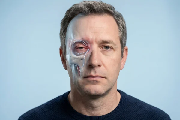
The aging process is a multifaceted biological phenomenon that extends far beyond the superficial layers of the epidermis. While public perception of aging is often dominated by the appearance of fine lines, wrinkles, and pigmentary changes, medical professionals are increasingly focusing on the profound structural shifts occurring beneath the surface. Dr. Steven Land, a prominent figure in aesthetic medicine, has highlighted facial bone resorption as a critical, yet often overlooked, driver of facial transformation. This physiological process involves the gradual loss of bone density and volume in the facial skeleton, fundamentally altering the scaffolding upon which soft tissues, muscles, and skin reside.
The Structural Foundation of Facial Aesthetics
To understand facial bone resorption, one must first view the face as a complex architectural system. The facial skeleton provides the essential framework that determines the projection of the cheeks, the definition of the jawline, and the support for the periorbital area. Unlike the long bones of the body, which primarily serve structural and locomotive functions, the bones of the face are intricately linked to the aesthetics of human expression and identity.
Bone is a dynamic tissue, constantly undergoing a process known as remodeling. This involves a delicate balance between osteoclasts, which break down bone tissue (resorption), and osteoblasts, which deposit new bone (ossification). During youth, these two processes are generally in equilibrium or favor bone deposition. However, as individuals age, the rate of resorption begins to outpace the rate of formation. This imbalance leads to a net loss of bone mass, a phenomenon that Dr. Land identifies as a primary cause of the "sagging" appearance often attributed solely to skin laxity.
The Biological Mechanism of Bone Remodeling
The pathophysiology of bone resorption is influenced by a combination of genetic, hormonal, and environmental factors. Hormonal shifts play a significant role, particularly in women. The decline in estrogen during perimenopause and menopause is a well-documented catalyst for systemic bone loss, including the facial skeleton. Estrogen is known to inhibit the activity of osteoclasts; therefore, its reduction accelerates the breakdown of bone matrix.
Furthermore, mechanical stress—or the lack thereof—impacts bone density. The "mechanostat" theory suggests that bone adapts its structure based on the physical loads placed upon it. In the face, the loss of teeth (edentulism) leads to a rapid resorption of the alveolar ridge of the mandible and maxilla because the bone no longer receives the mechanical stimulation of mastication. Even in individuals with full dentition, the natural atrophy of overlying facial muscles can reduce the mechanical load on the underlying bone, further contributing to the resorptive process.
Anatomical Chronology: How the Skeleton Shifts Over Time
Facial bone resorption does not occur uniformly across the skull; rather, it follows a specific and predictable pattern. Longitudinal studies using computed tomography (CT) imaging have allowed researchers to map these changes with high precision.
The Periorbital Region and Forehead
One of the earliest sites of resorption is the orbital rim. The eye sockets tend to widen and recede, particularly at the superomedial and inferolateral corners. This expansion of the orbital aperture causes the overlying soft tissue to lose support, leading to the appearance of "hollow" eyes, the protrusion of orbital fat (eye bags), and the descent of the eyebrows. Simultaneously, the frontal bone (forehead) loses convexity, flattening out and contributing to a more aged, tired appearance.
The Midface and Maxilla
The maxilla, or upper jawbone, undergoes significant retrusion with age. As the maxilla recedes, it provides less support for the overlying malar (cheek) fat pads. This lack of skeletal projection is a primary reason why the cheeks appear to "drop" and why the nasolabial folds (the lines from the nose to the corners of the mouth) become more pronounced. The pyriform aperture—the boney opening for the nose—also enlarges, which can cause the tip of the nose to lose support and appear to droop or lengthen.
The Lower Face and Mandible
The mandible, or lower jaw, is perhaps the most visible area of skeletal change. Resorption occurs along the body and the ramus of the jaw, leading to a loss of definition in the jawline. As the height and length of the mandible decrease, the chin may recede (microgenia), and the "angle of the jaw" becomes less obtuse. This loss of structural projection allows the soft tissues of the lower face to collapse into the neck, creating jowls and obscuring the distinction between the face and the throat.
Supporting Data and Clinical Insights
Quantitative data from aesthetic research journals underscores the severity of these changes. Studies comparing CT scans of younger individuals (ages 20–40) with older cohorts (60+) show significant decreases in the angles and dimensions of the facial skeleton. For instance, the maxillary angle decreases significantly with age, indicating a loss of midface projection.
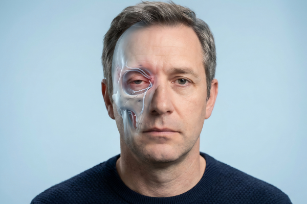
Research published in the Journal of Plastic and Reconstructive Surgery indicates that the most significant bone loss occurs in the midface and the jawline, with women often showing these changes earlier than men. Data suggests that by the age of 60, the average person may have lost up to 10-20% of their facial bone volume in specific high-impact areas. This volume loss creates a "surplus" of skin; because the underlying frame has shrunk, the skin appears too large for the face, leading to the characteristic folds and sags of the elderly.
Evolution of Aesthetic Interventions
The recognition of bone resorption has revolutionized the field of aesthetic medicine. In the late 20th century, the standard approach to facial rejuvenation was primarily surgical tightening—essentially pulling the skin tighter over a shrinking frame. While this addressed laxity, it often resulted in a "windblown" or unnatural appearance because it did not address the loss of volume.
In the current clinical landscape, practitioners like Dr. Steven Land advocate for a "pan-facial" approach that prioritizes structural restoration. This has led to the development of "deep-plane" injection techniques, where dermal fillers (such as hyaluronic acid or calcium hydroxylapatite) are placed directly onto the periosteum (the surface of the bone) to mimic the lost skeletal volume.
- Structural Volumization: By placing high-G-prime fillers in areas like the malar bone or the mandibular angle, clinicians can recreate the projection that was lost to resorption.
- Biostimulators: Treatments that stimulate the body’s own collagen and potentially influence bone health are being explored as long-term solutions.
- Surgical Implants: In cases of severe resorption, silicone or porous polyethylene implants may be used to permanently restore the chin, cheeks, or jawline.
Official Responses and Expert Consensus
The medical community has reached a broad consensus that aging is a three-dimensional process. The American Society of Plastic Surgeons (ASPS) and various aesthetic dermatology boards have updated their training modules to include detailed studies of craniofacial anatomy.
Experts emphasize that understanding the "why" behind the wrinkle is essential for patient safety and satisfaction. "If you only treat the skin without addressing the foundation, you are essentially trying to drape a tablecloth over a table that is missing its legs," noted one clinical educator in a recent symposium on regenerative aesthetics. The consensus is that a multi-layered approach—addressing bone, fat pads, muscle, and skin—is the only way to achieve natural-looking and long-lasting results.
Broader Impact and Implications
The implications of facial bone resorption extend beyond the cosmetic. It is an indicator of systemic health and bone density. There is an observed correlation between facial bone loss and osteoporosis. Dentists and maxillofacial surgeons often serve as the first line of defense, noticing changes in the jawbone that may suggest a need for systemic bone density screenings (DEXA scans).
Furthermore, the psychological impact of structural aging cannot be understated. Facial structure is central to human recognition and self-perception. As the skeletal foundation shifts, individuals often report a loss of "self" in the mirror. By addressing the root cause—the bone—aesthetic medicine provides more than just vanity; it provides a restoration of the patient’s original anatomical proportions.
Future Directions in Research
Looking ahead, the focus is shifting toward regenerative medicine. Scientists are investigating the use of growth factors and stem cell therapies to potentially slow or reverse bone resorption in the face. There is also significant interest in the role of nutrition—specifically Vitamin D, K2, and Calcium—and how optimized levels might preserve the facial skeleton into later life.
As imaging technology becomes more accessible, "prejuvenation" strategies are also evolving. Younger patients are being educated on the importance of maintaining bone health through lifestyle and preventative treatments, aiming to minimize the degree of resorption before it becomes clinically significant.
The insights provided by Dr. Steven Land and the wider medical community represent a paradigm shift. By acknowledging that the face "shrinks" as much as it "sags," the field of aesthetics has moved toward a more scientific, anatomically accurate model of care. Understanding facial bone resorption is no longer just a niche anatomical fact; it is the cornerstone of modern geriatric aesthetics and a vital component of our understanding of human biology.
