
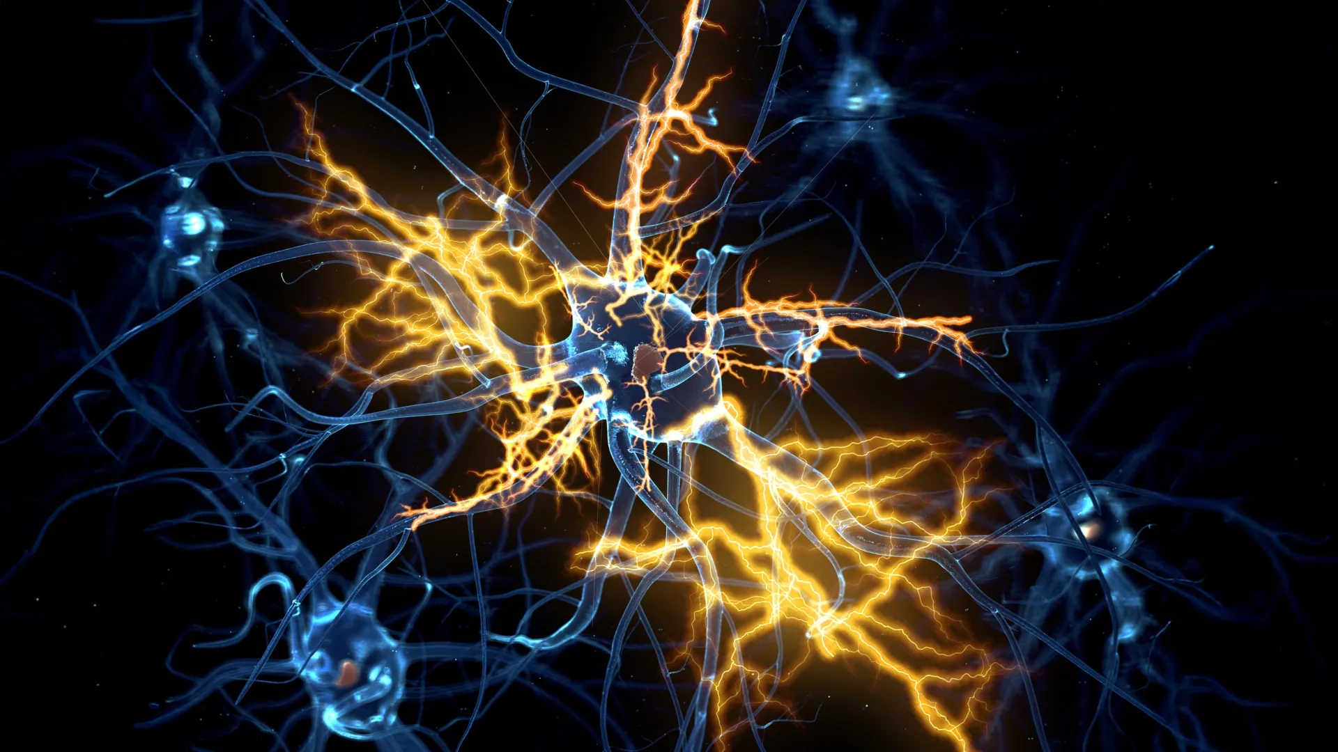
Scientists at the University of Cambridge have engineered sophisticated lab-grown brain and spinal cord systems, replicating the intricate pathways of human movement signals. This groundbreaking model has yielded a remarkable discovery: nerve damage, long considered irreversible, may indeed be amenable to repair under specific conditions, potentially revolutionizing treatment for debilitating neurological conditions.
Unraveling the Mysteries of Neural Regeneration
The journey from embryonic development to adulthood involves the intricate formation of communication networks between the brain and spinal cord. This complex architecture relies on axons, the long, slender projections of nerve cells, which act as vital conduits for transmitting signals that control everything from fine motor skills to essential bodily functions. In the early stages of human development, these axons possess a remarkable capacity for growth and regeneration. However, as the central nervous system matures, this regenerative ability significantly diminishes, leading to the often permanent consequences of injuries to the brain and spinal cord. This loss of regenerative potential is also a key factor in the progression of devastating neurological diseases such as motor neurone disease (MND) and multiple sclerosis (MS).
For decades, the prevailing scientific understanding held that once axons in the adult central nervous system were damaged, their capacity for regrowth was largely lost. This stark reality has contributed to the persistent and often devastating effects of spinal cord injuries, strokes, and neurodegenerative disorders, leaving millions worldwide facing paralysis, chronic pain, and loss of function. The limited success of regenerative therapies has underscored the urgent need for novel approaches that can overcome these intrinsic biological barriers.
Miniature Human Nervous Systems: A New Frontier in Research
Building upon their previous success in creating pea-sized "brain organoids" in 2021 – derived from patient stem cells and mimicking aspects of the cerebral cortex – Dr. András Lakatos and his team at the University of Cambridge have now advanced their research by constructing a miniature, interconnected human brain and spinal cord system. These sophisticated organoids, meticulously cultured in the lab, allow researchers to observe neural development and response to injury in a controlled environment that closely mirrors human physiology.
The innovative design of these new models addressed the anatomical separation of the brain and spinal cord. By maintaining the brain and spinal cord organoids in proximity but physically distinct within their culture, the researchers were able to witness axons from the brain tissue actively growing across the intervening gap to establish connections with the spinal cord tissue. This remarkable demonstration of directed axonal growth in vitro created a functional neural circuit, capable of triggering coordinated contractions in associated clusters of muscle cells – a crucial step in simulating how movement signals are transmitted and executed.
Developmental Timeline Reveals a Critical Window for Regeneration
The Cambridge team meticulously monitored these miniature nervous systems for over a year, a duration designed to capture the developmental trajectory of neural regeneration. Their observations revealed a critical period: up until approximately day 150 of development, a stage analogous to the middle trimester of pregnancy, damaged axons within these organoids retained a significant capacity for regrowth. Beyond this developmental milestone, however, the researchers observed a precipitous decline in the neurons’ regenerative capabilities.
George Gibbons, the lead author of the study from the Department of Clinical Neurosciences at the University of Cambridge, articulated the significance of these findings: "Neurons taken from less mature organoids regrew long fibers after injury, but those from more mature organoids showed a sharp drop in their ability to regrow. In other words, poor regeneration is built into human neurons as they mature in the central nervous system." This observation suggests that the cessation of axonal regeneration is not a sudden event but rather a programmed aspect of neural maturation.
Unlocking the Genetic Secrets of Axon Growth
To understand the underlying mechanisms driving this developmental shift, the researchers delved into the gene activity of the neurons connecting the brain and spinal cord. Their comprehensive analysis identified a complex network of genes that appears to function as a biological "switch," actively limiting axon growth as neurons mature and establish synaptic connections. This intricate genetic program, while essential for the orderly development of precise neural circuitry, inadvertently hinders the repair of damaged axons in adulthood.
In a pivotal experiment, the scientists demonstrated the plasticity of this system by selectively blocking key regulators within this identified gene network. The results were striking: the neurons, when released from these inhibitory genetic signals, regained their ability to grow axons, effectively reversing the developmental suppression of regeneration. This intervention offers a powerful proof-of-concept that the intrinsic limitations on nerve repair might be overcome through targeted molecular manipulation.
A Pharmaceutical Ally: Lynestrenol Shows Promise
The identification of this regulatory gene network opened up a new avenue for therapeutic exploration. The research team embarked on a systematic search through a comprehensive database of pharmaceutical compounds, seeking existing drugs that could modulate the activity of this newly discovered gene network. Among the promising candidates, lynestrenol emerged as a particularly noteworthy discovery. Lynestrenol is a synthetic progestogen, a type of hormone drug already approved for clinical use in managing certain menstrual disorders and as a contraceptive.
When lynestrenol was administered to the damaged neurons in the organoid models, it demonstrated a significant enhancement of axon regrowth. This finding is particularly encouraging as it suggests that a drug already in clinical use, with a known safety profile, could potentially be repurposed to promote nerve regeneration. While lynestrenol itself may not be the definitive solution for spinal cord repair, its efficacy in this model provides compelling evidence that directly targeting human neurons to stimulate axon regeneration is a viable therapeutic strategy.
Beyond Molecular Mechanisms: The Role of the Microenvironment
While the focus of this study was on intrinsic neuronal mechanisms, the researchers acknowledge that other factors can impede nerve repair in vivo. Scar tissue formation and inflammation are well-documented barriers to axonal regeneration following central nervous system injury. However, the Cambridge team emphasizes that understanding the neuron-specific biological processes that limit regeneration remains paramount. Their findings align with previous research indicating that younger neurons possess a greater ability to navigate and grow through environments that would typically inhibit repair in mature nervous systems. This suggests that by reactivating the inherent regenerative capacity of neurons, the challenges posed by the inhibitory microenvironment might be more effectively overcome.
A Paradigm Shift in Neurological Treatment
Senior author Dr. András Lakatos expressed profound optimism about the implications of their work: "When the brain and spinal cord are damaged, the nerve fibers that carry movement signals from the brain to the spinal cord rarely grow back. That’s why paralysis is usually permanent. But we didn’t know exactly when the ability of axons to regenerate becomes limited. Our model provides a good indication that this block happens during development, and it can still be reversed after this point."
He further elaborated on the potential of their findings: "Lynestrenol itself may not be the answer to spinal cord repair, but it shows us that, in principle, it should be possible to directly target human neurons and regenerate their axons. Although we still need to show that this strategy will also help to re-establish appropriate connections between the brain and spinal cord cells, this gives us hope that one day we may be able to treat conditions previously thought untreatable." The critical next step will involve demonstrating that the regenerated axons can form functional and appropriate connections within the complex neural circuitry, a prerequisite for restoring meaningful motor control.
The Growing Importance of Human Organoids in Medical Research
The development and application of organoid technology represent a significant leap forward in the study of human biology and disease. While animal models, such as rodents, have historically played a crucial role in neurological research, inherent biological differences can limit the direct translation of their findings to human patients. Human stem cell-derived organoids, like those developed by the Cambridge team, offer a more accurate recapitulation of human physiology, effectively bridging the gap between preclinical research and real-world patient outcomes.
Dr. Lakatos highlighted the unique value of these models: "Much of what we know about nerve regeneration comes from rodents, whose neurons behave differently from human neurons. Our sophisticated organoid models help bridge the knowledge gap from animal models to what we see in patients. They are also an important contribution to efforts to reduce the use of animals in research." This advancement not only accelerates the pace of discovery but also aligns with ethical imperatives to minimize animal testing in scientific endeavors.
The University of Cambridge is at the forefront of utilizing organoids across a broad spectrum of medical research, from investigating liver regeneration and the complexities of Crohn’s disease in pediatric populations to unraveling the earliest stages of human pregnancy. This burgeoning field promises to yield further transformative insights into a wide array of human health challenges.
The research was generously supported by grants from the UK Research and Innovation Medical Research Council and Spinal Research, underscoring the significant investment in this critical area of scientific inquiry. The findings offer a beacon of hope for individuals affected by conditions previously considered intractable, paving the way for the development of novel therapeutic strategies aimed at restoring function and improving quality of life.
