
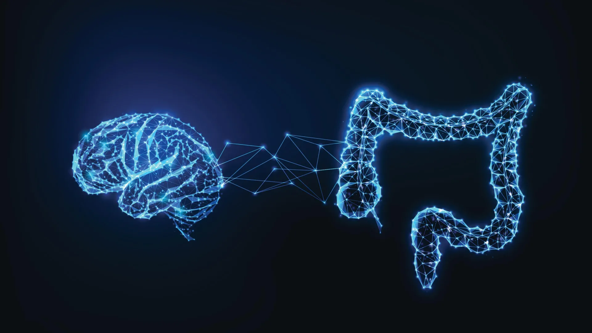
The taste of a madeleine famously plunged the French novelist Marcel Proust into a torrent of vivid childhood recollections, cementing the literary notion that flavor and memory are inextricably linked. For generations, this phenomenon was viewed exclusively through a neurological lens, attributing sensory-triggered nostalgia to the cerebral cortex and the hippocampus. However, groundbreaking new research indicates that the journey of a memory may begin far lower than the skull. Signals originating directly from the digestive system appear to play a decisive role in determining which food-related experiences are permanently encoded by the brain, redefining the intricate dialogue between our meals and our minds.
The study, published in the peer-reviewed journal Nature Communications, was led by Scott Kanoski, a professor of biological sciences at the USC Dornsife College of Letters, Arts and Sciences. The findings strongly suggest that the gastrointestinal tract actively contributes to memory formation, particularly regarding the crucial evolutionary task of locating and consuming sustenance. By examining the physiological pathways connecting the gut to the brain, researchers have uncovered a biological mechanism that prioritizes nutritional value over mere sensory gratification, while also shedding light on how modern diets may jeopardize long-term cognitive health.
Mapping the Gut-Brain Superhighway: The Vagus Nerve Connection
At the center of this discovery is the vagus nerve, one of the human body’s most critical neural conduits. Spanning from the brainstem down through the neck and chest to the abdomen, the vagus nerve acts as a bidirectional superhighway, constantly relaying status reports between internal organs and the central nervous system. While medical science has long understood that this nerve regulates visceral functions such as digestion, heart rate, and appetite, its direct involvement in higher cognitive processes like memory formation has remained largely unexplored until recently.
To decipher this communication route, Kanoski and his team conducted a series of controlled experiments utilizing animal models. The researchers discovered that when subjects consumed nutrient-dense foods, there was an immediate and substantial spike in the release of acetylcholine within the neurons of the hippocampus—the seahorse-shaped structure embedded deep within the temporal lobe that is universally recognized as the brain’s primary hub for learning and spatial memory.
Acetylcholine is a vital neurotransmitter that acts as a biochemical gatekeeper, facilitating synaptic plasticity and enabling the brain to record, retain, and retrieve new information. Crucially, the team observed that this acetylcholine surge was entirely dependent upon real-time physiological messages traveling upward from the gut via the vagus nerve. When the researchers surgically or chemically disrupted communication along this neural pathway, the post-meal spike in acetylcholine vanished. Consequently, the test subjects exhibited marked impairments in spatial memory tasks, struggling significantly to remember where they had recently encountered food sources.
Nutrients Versus Sweetness: The Brain Demands Substance
One of the most revealing aspects of the new study is the brain’s discerning ability to evaluate the biochemical composition of what we eat, distinguishing true nutritional value from superficial sensory traits. For decades, food scientists and psychologists have studied how sweetness and pleasant flavors trigger reward circuits in the brain, often leading to compulsive eating behaviors. Yet, the USC Dornsife findings demonstrate that the memory-encoding pathways operate on a fundamentally different principle.
During the experiments, subjects that consumed foods rich in fats and sugars displayed robust activation in memory-related neural circuits. However, when animals were given low-calorie or noncaloric liquids that mimicked the sweet taste of sugar without delivering actual metabolic energy, the expected memory-boosting response failed to materialize. The brain, it appears, is not easily fooled by artificial sweeteners or empty calories.
"We think the mechanism likely evolved to help animals remember vital information about food sources," explains Logan Lauer, a PhD student in Kanoski’s laboratory and the first author of the study. In a natural environment, where resources are scarce and unpredictable, remembering the exact geographic location of a reliable, energy-rich food source can mean the difference between life and death. When an animal forages and consumes a meal packed with vital calories and nutrients, the gut dispatches a chemical telegram to the brain essentially stating: "This meal provided valuable nutrients, so remember precisely where and how you obtained it."
Chronology of the Discovery and Methodology
The trajectory of this research reflects a growing shift in modern neuroscience toward the gut-brain axis, a field that has expanded exponentially over the past fifteen years. Initial hypotheses regarding the vagus nerve focused primarily on satiety and metabolic disorders, but advances in neural tracing and optogenetics allowed Kanoski’s team to observe real-time neurotransmitter fluctuations in response to specific macronutrients.
The research project spanned several years, beginning with foundational anatomical mappings of vagal afferent fibers connecting the gastrointestinal tract to the basal forebrain and hippocampus. Following these initial observations, the team designed behavioral and neurochemical assays to measure hippocampal acetylcholine levels during feeding events. By comparing subjects receiving caloric versus non-caloric stimuli, and subsequently replicating the tests following vagal deafferentation, the researchers isolated the vagus nerve as the indispensable conduit for nutrient-driven memory consolidation.
The collaborative effort behind the study brought together a multidisciplinary team of neuroscientists and biologists from USC Dornsife, alongside academic partners from Bucknell University and the Université de Montréal. Financial support for the extensive preclinical trials was provided by major grants from the National Institute of Diabetes and Digestive and Kidney Diseases, the National Institute on Aging, the Quebec Research Funds, and the Alzheimer’s Association.
The Cost of Convenience: How Unhealthy Diets Damage the Pathway
While the evolutionary purpose of the gut-brain memory pathway is to secure survival, modern dietary trends appear to short-circuit this delicate mechanism. The research team uncovered a troubling downside to frequent exposure to foods heavily laden with fats and refined sugars. Although such foods triggered strong short-term memory responses initially, prolonged and repeated consumption produced the exact opposite effect over time.
In subsequent trials, subjects raised on high-fat and high-sugar diets exhibited severely degraded communication between the gastrointestinal tract and the hippocampus. Most alarmingly, these deficits proved persistent; even after the subjects were transitioned back to a balanced, healthy diet, their memory-related brain responses remained permanently blunted. Behaviorally, these animals performed poorly on standardized cognitive evaluations designed to test spatial recall and environmental navigation.
These findings suggest that chronic ingestion of ultra-processed, highly caloric foods may physically erode or desensitize the very neurochemical systems designed to help us learn from our environment. The implications extend far beyond simple weight gain, pointing to a mechanism wherein poor dietary habits actively dismantle cognitive infrastructure.
Broader Implications for Public Health and Neurodegeneration
The intersection of diet, neurology, and cognitive decline has become one of the most urgent areas of medical research today. Epidemiological data has long established a clear correlation between the modern Western diet—characterized by high amounts of saturated fats and processed sugars—and an elevated risk of systemic metabolic disorders such as obesity and type 2 diabetes. Concurrently, these metabolic conditions are widely recognized as major independent risk factors for cognitive decline and dementia in aging populations.
The USC Dornsife study offers a tangible biological framework that bridges these clinical observations. If chronic exposure to unhealthy diets systematically degrades gut-to-brain communication, the resulting deficit in hippocampal acetylcholine signaling could serve as a foundational catalyst for broader cognitive deterioration.
Furthermore, this discovery opens intriguing new avenues for understanding and potentially treating neurodegenerative disorders such as Alzheimer’s disease. Dr. Kanoski highlights a critical clinical parallel: "The disruption of acetylcholine signaling in the hippocampus is one of the earliest neurochemical changes in Alzheimer’s disease." By demonstrating that this exact neurochemical system is naturally regulated and boosted by vagus nerve signaling from the gut, researchers are presented with an entirely novel therapeutic frontier.
Future Therapeutic Horizons and Vagus Nerve Stimulation
The possibility of leveraging the gut-brain axis for therapeutic gain has transformed from speculative science fiction into a viable medical pursuit. Vagus nerve stimulation (VNS)—a clinical procedure involving the surgical implantation of a device that delivers mild electrical impulses to the vagus nerve—is already FDA-approved and actively utilized in the management of drug-resistant epilepsy and treatment-resistant depression.
With the publication of these new findings, researchers are beginning to investigate whether similar neuromodulatory approaches could be adapted to support cognitive function, preserve memory, or mitigate the early stages of neurodegeneration. While the prospect of utilizing vagus nerve stimulation or targeted nutritional interventions to boost memory is still in its infancy, the conceptual shift is profound. Medical science may soon look past the confines of the cranium to treat cognitive decline, seeking solutions instead in the complex ecosystem of our digestive tracts.
Cautious Optimism and Next Steps
Despite the groundbreaking nature of the study, the research team emphasizes a necessary note of scientific caution. All primary experiments were conducted using animal models, and while mammalian biology shares deep evolutionary conservation regarding autonomic nervous systems, extensive clinical trials will be required to confirm whether identical gut-to-brain memory mechanisms operate in humans.
Nevertheless, the implications are vast. As researchers continue to untangle the biochemical dialogue between our plates and our minds, the old adage that "you are what you eat" takes on a startlingly literal neurological meaning. The next time a familiar flavor transports a diner across decades of memory, they may have more than just their brain to thank—they may owe an equal debt to the quiet, complex machinery of the gut.
