
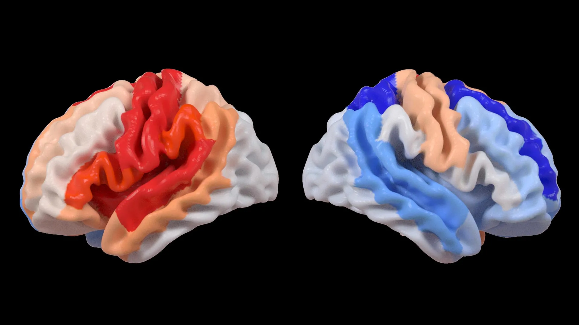
A groundbreaking study published in the esteemed journal The Lancet Digital Health has unveiled a remarkable and previously underestimated capacity of the human brain to adapt following a stroke. Researchers at the USC Mark and Mary Stevens Neuroimaging and Informatics Institute (Stevens INI) have identified a phenomenon where individuals experiencing significant physical limitations after a stroke exhibit signs of a “younger” brain structure in areas spared from the initial injury. This intriguing finding suggests a profound level of brain reorganization and compensatory rewiring that occurs as the brain attempts to regain lost function.
The research, a collaborative effort under the umbrella of the Enhancing NeuroImaging Genetics through Meta-Analysis (ENIGMA) Stroke Recovery Working Group, represents a significant leap forward in understanding the intricate aftermath of cerebrovascular incidents. The scientists meticulously analyzed a vast dataset comprising brain scans from over 500 stroke survivors. These crucial data points were collected across 34 distinct research centers spanning eight countries, a testament to the global scope and ambition of this research initiative. By employing sophisticated deep learning models, trained on an immense repository of tens of thousands of Magnetic Resonance Imaging (MRI) scans, the team was able to estimate the “brain age” of various regions within each cerebral hemisphere. This novel approach allowed them to examine not only the immediate structural impact of stroke but also its influence on the brain’s subsequent recovery processes.
AI Unveils a Younger Brain on the Opposite Side of Injury
At the heart of this discovery lies the application of advanced artificial intelligence, specifically a type known as a graph convolutional network. This powerful AI system was instrumental in estimating the biological age of 18 specific brain regions based on the detailed anatomical information provided by the MRI scans. This estimated biological age was then compared to each participant’s chronological age, yielding a metric termed the “brain-predicted age difference” (brain-PAD). A positive brain-PAD, indicating a younger predicted age than actual, is generally associated with better brain health, while a negative brain-PAD suggests accelerated aging.
The most striking revelation emerged when these brain age measurements were correlated with motor function scores. Stroke survivors who experienced severe impairments in movement, even after more than six months of intensive rehabilitation, displayed a younger-than-expected brain age in regions located in the hemisphere opposite to the site of the stroke. This effect was particularly pronounced in the frontoparietal network, a critical brain circuitry responsible for a complex array of functions including movement planning, attentional control, and the coordination of various cognitive and motor processes.
Dr. Hosung Kim, an associate professor of research neurology at the Keck School of Medicine of USC and a co-senior author of the study, elaborated on the implications of these findings. “We discovered that larger strokes tend to accelerate aging in the damaged hemisphere,” Dr. Kim stated. “However, paradoxically, they appear to make the opposite side of the brain seem younger. This pattern strongly suggests that the brain is actively reorganizing itself, essentially rejuvenating undamaged networks to compensate for the functions that have been lost.”
The Mechanism of Compensatory Neuroplasticity
This observed phenomenon offers a compelling glimpse into the brain’s remarkable neuroplasticity – its inherent ability to reorganize its structure, function, and connections throughout life. In the context of stroke, where neuronal damage can be extensive and irreversible, the brain appears to initiate a strategic redistribution of resources. The undamaged hemisphere, particularly specific networks like the frontoparietal network, seems to step up its activity and potentially undergoes structural adaptations to shoulder some of the cognitive and motor demands previously handled by the lesioned areas.
“These findings suggest that when stroke damage leads to greater movement loss, undamaged regions on the opposite side of the brain may adapt to help compensate,” Dr. Kim explained. “We observed this particularly in the contralesional frontoparietal network, which exhibited a more ‘youthful’ pattern and is known to support motor planning, attention, and coordination. Rather than indicating a complete recovery of movement, this pattern may actually reflect the brain’s intricate attempt to adjust and function when the damaged motor system can no longer operate normally. This provides us with an entirely new perspective on neuroplasticity, one that traditional imaging techniques might not have been able to capture.”
The frontoparietal network, often referred to as the dorsal attention network, plays a pivotal role in directing attention to relevant stimuli and is deeply involved in the executive control of voluntary movements. Its involvement in this compensatory mechanism underscores the brain’s sophisticated strategies for maintaining functional integrity in the face of severe injury. The observed “rejuvenation” in these areas, as indicated by the brain age prediction, could signify increased neuronal density, enhanced synaptic plasticity, or more efficient neural signaling within these undamaged circuits, all aimed at mitigating the deficits caused by the stroke.
The Power of Global Collaboration and Big Data
The success of this ambitious study hinges significantly on the ENIGMA consortium, a global collaborative initiative that aggregates neuroimaging and genetic data from over 50 countries. ENIGMA’s mission is to advance our understanding of the brain in both health and disease by creating unprecedentedly large and diverse datasets. By standardizing MRI data acquisition protocols and clinical information from a multitude of research groups worldwide, the team successfully assembled the most extensive stroke neuroimaging dataset of its kind to date. This large-scale approach is crucial for detecting subtle patterns that might be missed in smaller, single-institution studies.
Arthur W. Toga, PhD, director of the Stevens INI and Provost Professor at USC, highlighted the transformative impact of this collaborative model. “By pooling data from hundreds of stroke survivors globally and applying cutting-edge artificial intelligence, we are able to detect subtle patterns of brain reorganization that would remain invisible in smaller studies,” Dr. Toga stated. “These findings, revealing regionally differential brain aging in chronic stroke, hold the potential to eventually guide the development of highly personalized rehabilitation strategies.”
The sheer volume of data analyzed in this study, coupled with the advanced AI algorithms, allowed researchers to move beyond broad observations and identify nuanced changes at a regional level. The consistent observation of a younger brain age in the contralesional frontoparietal network across a diverse cohort of stroke survivors strengthens the evidence for this adaptive mechanism. This suggests that the brain’s response to stroke is not uniform but exhibits specific patterns of regional activation and potential rejuvenation.
Timeline of Discovery and Future Directions
The journey to these findings has been a multi-year endeavor, building upon decades of research into stroke recovery and the development of sophisticated neuroimaging techniques. The ENIGMA consortium itself has been instrumental in fostering international collaboration for over a decade, facilitating the large-scale data sharing necessary for such impactful research. The specific methodology employed in this study, utilizing deep learning for brain age prediction, represents a more recent advancement, likely building on earlier work in machine learning applied to neuroimaging.
The current study focused on chronic stroke survivors, typically defined as those more than six months post-stroke, to capture the long-term adaptive changes in the brain. However, the researchers are keen to extend their investigations to observe these patterns in real-time. Future research plans include longitudinally tracking patients from the acute stages following a stroke through their long-term recovery. By monitoring how brain aging patterns and structural changes evolve over time, clinicians could gain invaluable insights into an individual’s unique recovery trajectory.
This longitudinal approach is expected to provide a dynamic understanding of neuroplasticity, revealing whether the observed “younger” brain age in certain regions is a stable compensatory mechanism or a sign of ongoing adaptation. Such knowledge could pave the way for precisely tailored rehabilitation programs, optimizing interventions based on an individual’s specific neural response to injury. The ultimate goal is to enhance recovery outcomes and significantly improve the quality of life for stroke survivors worldwide.
Implications for Personalized Stroke Rehabilitation
The implications of this research extend far beyond academic curiosity, holding tangible promise for the future of stroke rehabilitation. The ability to identify specific brain regions that are adapting to compensate for injury, and to quantify this adaptation through brain age metrics, could revolutionize how therapy is delivered.
Currently, stroke rehabilitation often follows standardized protocols, which may not be optimal for every patient given the vast heterogeneity in stroke types, locations, and individual responses. This new understanding suggests that personalized interventions could be developed by identifying which compensatory networks are most active and potentially most amenable to enhancement. For example, therapies could be designed to specifically bolster the function of the contralesional frontoparietal network in patients showing pronounced signs of its rejuvenation, thereby maximizing its potential to support motor control and coordination.
Furthermore, the brain age metric could serve as a prognostic tool. A more pronounced “younger” brain age in compensatory areas might correlate with a better capacity for functional recovery, while a lack of such changes could indicate a need for more intensive or different therapeutic approaches. This data-driven approach could lead to more efficient use of rehabilitation resources and a more predictable recovery pathway for patients and their caregivers.
A New Window into Neuroplasticity
The study’s findings offer a novel and powerful way to visualize and quantify neuroplasticity, a concept that has long been central to understanding brain recovery but has been challenging to measure objectively and regionally. Traditional imaging techniques, while invaluable, often focus on gross structural changes or functional activation patterns. The brain age prediction method, powered by AI, provides a more nuanced assessment of the biological state of brain tissue and its functional potential.
This method effectively bridges the gap between structural and functional assessments by providing an integrated measure of brain health. The identification of regionally differential brain aging suggests that the brain’s response to stroke is complex, with some areas showing signs of accelerated aging due to damage, while others exhibit a compensatory “youthfulness.” This duality is a critical insight into the intricate interplay of degeneration and adaptation post-stroke.
Broader Impact and Support for the Research
The study, titled "Deep learning prediction of MRI-based regional brain age reveals contralesional neuroplasticity associated with severe motor impairment in chronic stroke: A worldwide ENIGMA study," received crucial funding from the National Institutes of Health (NIH) under grant R01 NS115845. This significant investment underscores the importance placed on advancing stroke research by national health organizations. The project also benefited from the invaluable support of international collaborators from leading institutions, including the University of British Columbia, Monash University, Emory University, and the University of Oslo, further highlighting the global commitment to tackling the challenges of stroke recovery.
The implications of this research are far-reaching, potentially impacting not only stroke survivors but also informing our understanding of brain adaptation in other neurological conditions. The development and validation of AI-driven methods for assessing brain health and plasticity hold immense promise for the future of neurological care, moving towards more precise, personalized, and effective interventions. The ongoing work by the Stevens INI, including the creation of educational materials like the explanatory video on contralesional neuroplasticity, demonstrates a commitment to disseminating these vital findings to both the scientific community and the public.
This comprehensive study, with its innovative methodology, global reach, and profound findings, marks a significant milestone in stroke research. It opens new avenues for understanding the brain’s remarkable resilience and offers a beacon of hope for improved rehabilitation strategies and better outcomes for millions affected by stroke each year. The continued exploration of these AI-driven insights promises to reshape our approach to neurological recovery and enhance the lives of individuals worldwide.
