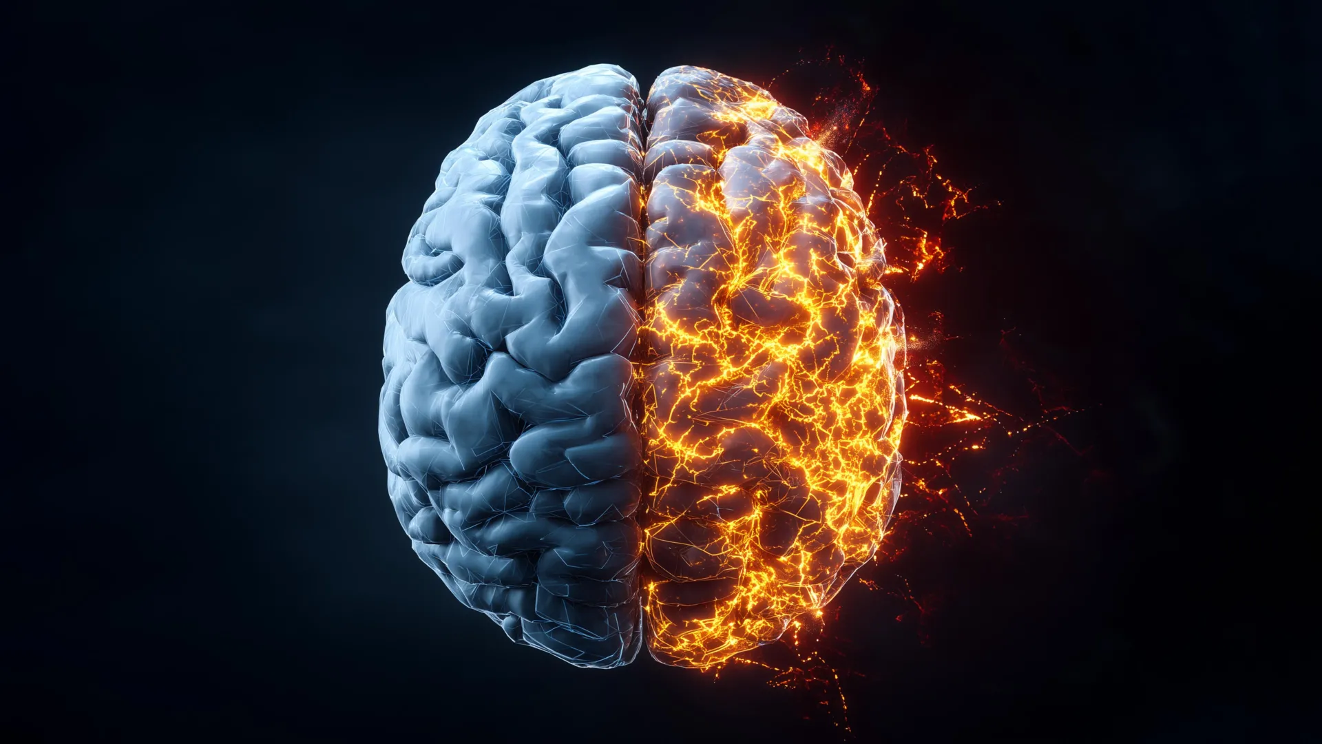
Researchers at Rice University have achieved a significant breakthrough in understanding Alzheimer’s disease, producing the first comprehensive, label-free molecular atlas of the disease in an animal model. This pioneering work offers an unprecedentedly deep look into the intricate mechanisms by which Alzheimer’s begins and progresses, a crucial step forward in combating a devastating illness that claims more lives annually than breast and prostate cancers combined. The findings, recently published in the prestigious journal ACS Applied Materials and Interfaces, illuminate the complex and uneven distribution of chemical changes throughout the brain, challenging previous assumptions about the localized nature of Alzheimer’s pathology.
Illuminating the Brain’s Molecular Landscape: A Novel Imaging Approach
The Rice University team’s innovative approach hinges on the sophisticated integration of an advanced light-based imaging technique with cutting-edge machine learning algorithms. By meticulously examining brain tissue from both healthy and Alzheimer’s-affected animal models, they were able to visualize and quantify subtle molecular alterations that have eluded detection by conventional methods. This advanced methodology allowed them to move beyond the traditional focus on amyloid plaques, revealing a more pervasive and intricate pattern of chemical shifts across the entire brain.
At the heart of this discovery lies hyperspectral Raman imaging, a powerful evolution of Raman spectroscopy. This technique employs a precisely tuned laser to probe tissue, eliciting unique "chemical fingerprints" from individual molecules. Unlike traditional Raman spectroscopy, which provides a single data point of chemical information at a specific molecular site, hyperspectral Raman imaging performs this analysis thousands of times across an entire tissue slice. This exhaustive data collection process generates a high-resolution map, detailing the spatial variations in chemical composition throughout different brain regions.
"Traditional Raman spectroscopy takes one measurement of chemical information per molecular site," explained Ziyang Wang, an electrical and computer engineering doctoral student at Rice and a first author of the study. "Hyperspectral Raman imaging repeats this measurement thousands of times across an entire tissue slice to build a full map. The result is a detailed picture showing how chemical composition varies across different regions of the brain."
The researchers meticulously scanned entire brains, section by section, compiling an immense number of overlapping measurements to construct these high-resolution molecular maps. A critical aspect of their methodology is that the imaging process is "label-free." This means that the brain tissue samples were not subjected to treatment with dyes, fluorescent proteins, or other molecular tags that can sometimes alter the native state of the tissue.
"This means we observed the brain as is, capturing a complete, unaltered portrait of its chemical makeup," Wang elaborated. "I think this makes the approach more unbiased and better suited for discovering new disease-related changes that might otherwise be missed." This label-free approach is particularly valuable in the study of complex diseases like Alzheimer’s, where the introduction of exogenous labels could potentially interfere with or mask subtle, endogenous molecular changes.
Unraveling the Uneven Tapestry of Alzheimer’s Damage Through Machine Learning
The sheer volume of data generated by the hyperspectral Raman imaging process presented a significant analytical challenge. To address this, the research team turned to the power of machine learning (ML). Their analytical strategy involved a two-pronged approach, beginning with unsupervised ML algorithms. These algorithms were designed to identify natural patterns within the vast chemical data without any preconceived notions or prior assumptions about the expected outcomes. This initial phase allowed the ML models to independently group and sort tissue samples based purely on their intrinsic molecular characteristics.
Following this unsupervised exploration, the researchers employed supervised ML. In this stage, the trained models were fed specific examples of both Alzheimer’s-affected and healthy brain tissue. This training enabled the algorithms to learn the distinct molecular signatures associated with the disease and to accurately classify new samples. Crucially, this supervised learning phase allowed the researchers to quantify the degree to which different brain regions exhibited Alzheimer’s-related chemical alterations.
"We found that the changes caused by Alzheimer’s disease are not spread evenly across the brain," Wang stated. "Some regions show strong chemical changes, while others are less affected. This uneven pattern helps explain why symptoms appear gradually and why treatments that focus on only one problem have had limited success." This finding is particularly significant as it provides a molecular basis for the heterogeneous presentation of Alzheimer’s symptoms and the challenges in developing effective, universally targeted therapies.
Metabolic Disturbances: A Deeper Dive into Brain Function
Beyond the well-documented accumulation of aberrant proteins like amyloid-beta and tau, the study uncovered evidence of broader metabolic disruptions in the brains of Alzheimer’s-affected animals. The molecular atlas revealed significant variations in the levels of cholesterol and glycogen across different brain regions when compared to healthy controls. These metabolic shifts were most pronounced in areas critically involved in memory formation and recall, specifically the hippocampus and the cerebral cortex.
Cholesterol plays a vital role in maintaining the structural integrity and fluidity of neuronal cell membranes, essential for proper brain function and communication. Glycogen, on the other hand, serves as a readily accessible local energy reserve for brain cells. The observed depletion or alteration of these key metabolic components in memory-associated regions suggests that Alzheimer’s disease impacts not only the structural components of neurons but also their fundamental energy supply and maintenance mechanisms.
"Cholesterol is important for maintaining brain cell structure, and glycogen serves as a local energy reserve," commented Shengxi Huang, an associate professor of electrical and computer engineering and materials science and nanoengineering at Rice, and the corresponding author of the study. "Together, these findings support the idea that Alzheimer’s involves broader disruptions in brain structure and energy balance, not only protein buildup and misfolding." Professor Huang is also an affiliated member of several esteemed research institutes at Rice, including the Ken Kennedy Institute, the Rice Advanced Materials Institute, and the Smalley-Curl Institute, underscoring the interdisciplinary nature of this research.
A Chronological Journey: From Small-Scale Measurements to a Global Brain View
The genesis of this groundbreaking project can be traced back to ongoing discussions within the research community about novel approaches to unraveling the complexities of the Alzheimer’s brain. Initially, the team’s investigations were focused on analyzing relatively small, localized areas of brain tissue. However, a pivotal moment in the research occurred when Wang proposed a more ambitious goal: to map the entire brain.
"At first, we were measuring only small areas of brain tissue," Wang recounted. "Then I thought, what if we could map the entire brain and gain a much broader view? It took several rounds of testing and trial and error before the measurements and analysis worked well together." This iterative process involved refining the hyperspectral Raman imaging parameters to ensure optimal data acquisition across large tissue volumes and developing robust analytical pipelines to handle the massive datasets.
The successful integration of the imaging and machine learning components marked a turning point. When the complete chemical map of the brain finally coalesced, the impact was immediate and profound. New patterns, previously obscured by the limitations of conventional imaging techniques, began to emerge.
"Patterns emerged that had not been visible under regular imaging," Wang expressed with satisfaction. "Seeing those results was deeply satisfying. It felt like revealing a hidden layer of information that had been there all along, waiting for the right way to be analyzed." This moment underscores the transformative power of innovative technological development in scientific discovery, allowing researchers to perceive biological phenomena in entirely new ways.
Implications and Future Directions: Towards Earlier Diagnosis and Targeted Therapies
The delivery of the first detailed, dye-free chemical maps of the Alzheimer’s brain represents a significant advancement, offering a more holistic and nuanced understanding of the disease’s progression. This comprehensive molecular atlas provides a richer context for interpreting the pathological processes at play, moving beyond a singular focus on protein aggregation.
The implications of this research are far-reaching. By identifying specific regional vulnerabilities and metabolic dysfunctions, the findings could pave the way for earlier and more accurate diagnosis of Alzheimer’s disease. Current diagnostic methods often rely on the detection of symptoms that appear when significant neuronal damage has already occurred. A detailed molecular map could potentially reveal the earliest molecular signatures of the disease, allowing for intervention at a much earlier stage, when treatments are likely to be more effective.
Furthermore, the revelation of uneven disease progression and the identification of diverse molecular alterations across different brain regions have critical implications for therapeutic development. The limited success of previous treatments may be partly attributable to their broad, non-specific mechanisms or their focus on only one aspect of a multifaceted disease. This new atlas provides a roadmap for developing more targeted therapies that can address specific molecular pathways or regional vulnerabilities implicated in Alzheimer’s.
The research team at Rice University is hopeful that their work will serve as a foundational resource for the broader scientific community. By providing an unprecedented level of detail about the molecular landscape of the Alzheimer’s brain, this study empowers researchers to ask new questions and to design more effective strategies for slowing or even halting the progression of this debilitating condition. The ability to visualize and analyze the brain’s chemical composition without labels promises to accelerate discoveries in neurodegenerative diseases and beyond.
The research was generously supported by funding from several prominent organizations, including the National Science Foundation (grants 2246564 and 1934977), the National Institutes of Health (grant 1R01AG077016), and the Welch Foundation (grant C2144). This multi-agency support highlights the recognized importance and potential impact of this pioneering work in the fight against Alzheimer’s disease.