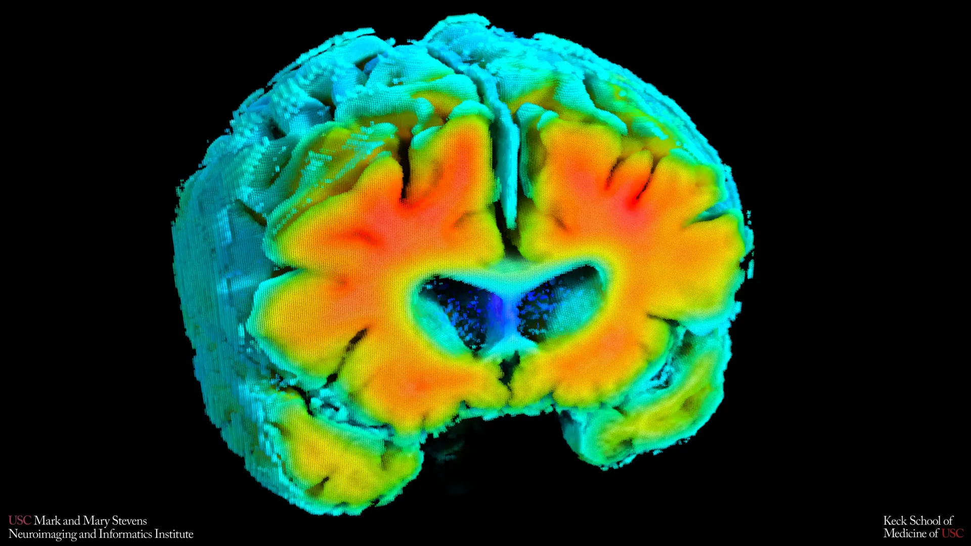
Small shifts in how blood moves through the brain and how brain cells receive oxygen may be closely connected to the risk of Alzheimer’s disease, according to groundbreaking new research from the Mark and Mary Stevens Neuroimaging and Informatics Institute (Stevens INI) at the Keck School of Medicine of USC. This pioneering study, published in the prestigious journal Alzheimer’s and Dementia: The Journal of the Alzheimer’s Association, utilized noninvasive techniques to uncover subtle yet significant correlations between cerebrovascular health and early markers of Alzheimer’s.
The research team, led by PhD candidate Amaryllis A. Tsiknia and senior author Meredith N. Braskie, PhD, examined a cohort of older adults, encompassing both individuals with and without cognitive impairment. Their findings suggest that the efficiency of the brain’s circulatory system, specifically blood flow velocity and oxygen saturation, can serve as an early indicator of Alzheimer’s risk, potentially predating the onset of noticeable cognitive decline.
Unveiling the Vascular Connection in Alzheimer’s
For decades, the scientific community has primarily focused on amyloid-beta plaques and tau tangles as the principal culprits in the pathogenesis of Alzheimer’s disease. However, this latest research from USC’s Stevens INI underscores the critical, and perhaps underestimated, role of vascular health in the disease’s progression.
"Amyloid and tau are often considered the primary players in Alzheimer’s disease, but blood flow and oxygen delivery are also critical," stated Amaryllis A. Tsiknia, the lead author of the study and a USC PhD candidate. "Our results show that when the brain’s vascular system functions more like it does in healthy aging, we also see brain features that are linked to better cognitive health."
The study’s implications are far-reaching, suggesting a paradigm shift in how Alzheimer’s might be detected and potentially managed in its nascent stages. By focusing on the intricate dance between blood circulation and cellular oxygenation, researchers are opening new avenues for early intervention and the development of preventative strategies.
Innovative Noninvasive Tools for Assessing Brain Circulation
A key strength of this research lies in its innovative use of noninvasive methodologies to quantify brain blood flow and oxygenation. The researchers employed two painless techniques that require participants to simply rest quietly:
- Transcranial Doppler Ultrasound (TCD): This established imaging technique uses sound waves to measure the speed at which blood travels through the major arteries supplying the brain. Faster blood flow in specific arteries can indicate the brain’s compensatory mechanisms in response to changes in blood pressure or carbon dioxide levels, reflecting the vascular system’s responsiveness.
- Near-Infrared Spectroscopy (NIRS): NIRS utilizes light waves to assess how effectively oxygen is delivered to the brain tissue, particularly the cerebral cortex, which is located near the surface of the brain. This method provides crucial insights into the oxygenation status of brain cells.
By integrating data from these two modalities, the research team developed sophisticated mathematical models to derive comprehensive indicators of cerebrovascular function. These indicators effectively capture how well the brain’s vascular network adapts to physiological fluctuations, such as changes in blood pressure and carbon dioxide levels, which are vital for maintaining optimal brain function. This nuanced approach moves beyond simple flow measurements to assess the dynamic regulatory capacity of the brain’s blood vessels.
Vascular Health as a Predictor of Alzheimer’s Biomarkers
The study’s most compelling findings emerged when these cerebrovascular indicators were correlated with established Alzheimer’s biomarkers. Participants exhibiting vascular function profiles more closely aligned with those of cognitively healthy individuals consistently demonstrated:
- Lower levels of amyloid plaque buildup: Amyloid plaques are abnormal protein deposits that accumulate in the brain and are a hallmark of Alzheimer’s disease, contributing to neuronal damage.
- Larger hippocampal volume: The hippocampus, a critical region of the brain responsible for memory formation and retrieval, often shrinks in individuals with Alzheimer’s. A larger hippocampus is associated with better memory function and reduced risk of cognitive decline.
These correlations are significant because they link the health of the brain’s blood vessels directly to the pathological processes and structural changes characteristic of Alzheimer’s disease. It suggests that a well-functioning vascular system may play a protective role, potentially mitigating the accumulation of toxic proteins and preserving vital brain structures.
"These vascular measures are capturing something meaningful about brain health," commented Dr. Meredith N. Braskie, senior author of the study and an assistant professor of neurology at the Keck School of Medicine. "They appear to align with what we see on MRI and PET scans that are commonly used to study Alzheimer’s disease, providing important information about how vascular health and standard brain measures of Alzheimer’s disease risk may be related."
Supporting Evidence from Mild Cognitive Impairment and Dementia
Further bolstering the study’s conclusions, the researchers observed a clear distinction in vascular function between cognitively normal participants and those diagnosed with mild cognitive impairment (MCI) or dementia. Individuals with MCI or dementia exhibited demonstrably weaker cerebrovascular function compared to their cognitively healthy counterparts.
This observation strongly supports the growing consensus that Alzheimer’s disease is not solely a neurodegenerative disorder but rather a complex condition with significant vascular contributions. The decline in blood vessel health appears to be an integral part of the broader Alzheimer’s disease continuum, potentially acting as an early trigger or accelerator of neurodegenerative processes.
Dr. Arthur W. Toga, director of the Stevens INI, emphasized the significance of these findings: "These findings add to growing evidence that Alzheimer’s involves meaningful vascular contributions in addition to classic neurodegenerative changes. Understanding how blood flow and oxygen regulation interact with amyloid and brain structure opens new doors for early detection and potentially prevention."
The Timeline of Alzheimer’s Research and Vascular Focus
The exploration of vascular contributions to cognitive decline has a history spanning several decades, but recent advancements in neuroimaging and data analysis have significantly accelerated this area of research. Early studies in the mid-20th century began to identify links between cardiovascular risk factors, such as hypertension and diabetes, and an increased likelihood of cognitive impairment in later life. However, distinguishing the direct impact of vascular disease from other age-related changes proved challenging.
The advent of advanced neuroimaging techniques like MRI and PET scans in the late 20th and early 21st centuries allowed researchers to visualize amyloid plaques and tau tangles with greater precision, solidifying their central role in Alzheimer’s pathology. Simultaneously, improvements in Doppler ultrasound and NIRS technology have enabled more detailed and accessible assessments of brain blood flow and oxygenation.
The USC study, published in 2023, represents a culmination of these technological advancements, providing robust, noninvasive evidence that integrates vascular markers with established Alzheimer’s pathology. It builds upon prior research from institutions like the Alzheimer’s Disease Neuroimaging Initiative (ADNI), which has consistently highlighted the importance of vascular risk factors in Alzheimer’s disease progression and the effectiveness of certain interventions. For example, studies have shown that managing blood pressure can reduce the risk of cognitive decline, suggesting a direct link between cardiovascular health and brain health.
Implications for Early Detection and Broader Screening
The noninvasive nature of the tools employed in this study offers a significant advantage for widespread application. Compared to MRI and PET imaging, transcranial Doppler ultrasound and near-infrared spectroscopy are:
- Less costly: Reducing the financial barrier to access for screening and research.
- Easier to perform: Requiring less specialized equipment and personnel.
- Patient-friendly: They do not involve injections of contrast agents, exposure to radiation, or demanding cognitive tasks, making them suitable for a broader range of individuals, including those with limited mobility or who are claustrophobic.
This simplicity could pave the way for large-scale screening programs, allowing for earlier identification of individuals at higher risk for Alzheimer’s disease. Such early detection is crucial, as interventions are generally more effective when initiated before significant neuronal damage has occurred.
The authors acknowledge that their findings represent a snapshot in time and do not definitively establish a cause-and-effect relationship. However, ongoing longitudinal studies are currently tracking participants to determine whether changes in these vascular measures can accurately predict future cognitive decline or predict response to potential treatments.
"If we can track these signals over time, we may be able to identify people at higher risk earlier and test whether improving vascular health can slow or reduce Alzheimer’s-related brain changes," Tsiknia added, highlighting the forward-looking potential of this research.
Future Directions and Potential Interventions
The findings from the Stevens INI study open up exciting new avenues for therapeutic development. If compromised cerebrovascular function is indeed an early contributor to Alzheimer’s, then interventions aimed at improving vascular health could become a critical component of Alzheimer’s prevention and treatment strategies. This could include:
- Lifestyle modifications: Encouraging healthy diets, regular exercise, and smoking cessation, all of which have proven benefits for cardiovascular health.
- Pharmacological interventions: Exploring existing medications for hypertension, cholesterol management, and diabetes that might also have neuroprotective effects.
- Novel therapeutic targets: Developing new drugs specifically designed to enhance blood flow, improve oxygen delivery, or repair damaged blood vessels in the brain.
Furthermore, the ability to monitor cerebrovascular function noninvasively could allow researchers to objectively assess the effectiveness of these interventions in real-time, accelerating the pace of clinical trials and drug development.
About the Study and Funding
The research was conducted by a dedicated team of scientists at the Keck School of Medicine of USC. In addition to Amaryllis A. Tsiknia and Meredith N. Braskie, the study’s other authors include Peter S. Conti, Rebecca J. Lepping, Brendan J. Kelley, Rong Zhang, Sandra A. Billinger, Helena C. Chui, and Vasilis Z. Marmarelis.
This groundbreaking work was made possible through significant funding from federal agencies dedicated to advancing scientific understanding and combating disease. The research received support from the Office of The Director, National Institutes of Health, under Award Number S10OD032285, and from the National Institute on Aging (NIA) through grant R01AG058162. This financial backing underscores the national recognition of the importance and potential impact of this research in the fight against Alzheimer’s disease.